
Page 29 - Read Online
P. 29
Page 8 of 16 Merhi et al. J Cancer Metastasis Treat 2021;7:42 https://dx.doi.org/10.20517/2394-4722.2021.80
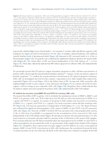
Figure 3. HF induces resistance to apoptosis in AML cells. (A) Representative cytograms of AML blood cells treated for 72 h with HF
(1.4, 2, and 3 µg/mL); detection of dead cells after annexin-V-FITC/PI staining and flow cytometry. The percentage of annexin-V-
positive cells is shown. (B) Light microscopy of non-treated and AML cells treated with HF: original magnification, 600×; May-
Grünwald stain; Scale bar, 10 µm. (C) Representative cytograms of PBMCs and isolated monocytes from two healthy donors, treated
with 2 µg/mL HF for 72 h, and cell death was assessed as described in (A). (D) Representative cytograms of AML cells isolated from
blood and bone marrow of one AML patient, treated with 2 µg/mL HF for 72 h, and cell death was assessed as described in (A). (E) Cell
death levels (2 µg/mL, 72 h) were determined in untreated and HF-treated malignant cells from 35 AML patients. Mean concentrations
± SD are indicated; P value was calculated using a Mann-Whitney U-test; ****P < 0.0001. (F) The percentage of HF-mediated AML cell
death was determined for all FAB subtypes by subtracting the percentage of annexin-V-positive cells in the absence of HF from the
percentage of annexin-positive cells in the presence of HF, and then dividing by the percentage of annexin-positive cells in the presence
of HF × 100. Data are the mean ± SEM (M0, n = 3; M1, n = 8; M2, n = 14; M3, n = 1; M4, n = 5; and M5, n = 4). P values were calculated
using one-way ANOVA (without M3 value); ns: not significant. (G&H) AML cells were treated with HF (2 µg/mL, 72 h) and the
percentage of annexin-V-positive cells was determined as described in (A) (left). In parallel, active caspase-3 expression was measured
by flow cytometry (right); cells were stained with FITC-rabbit Ig (control, dashed line) or anti-active caspase-3-FITC (solid line); the
percentages refer to the percentage of active caspase-3. (G) Responder AML cells are sensitive to HF treatment with death induction
(38% vs. 11% for untreated) and caspase-3 activation (34%). (H) Non-responder AML cells do not respond to HF treatment (no cell
death and no caspase-3 activation).
respectively, exhibited high levels of basal death (> 70% annexin-V-positive cells) and did not respond to HF
treatment [see Figure 3H (left) for M4 patient]. For the other 35 samples, cultured leukemic cells exhibited
variable baseline levels of spontaneous death [Figure 3E]. Exposure to HF increased death in 33 of the 35
blood samples [Figure 3E]. The paired-t test confirmed the significant enhanced death in HF-treated AML
cells [Figure 3E]. The lethal effect of HF was found independent of the FAB subtype (P = 0.0735)
[Figure 3F]. These data collectively indicate that HF induces apoptosis in cultured AML cells independently
of FAB status.
We previously showed that HF induced caspase-dependent apoptosis in AML cell lines representative of
primary AML cells through the mitochondrial (intrinsic) pathway . Caspase-3 is the executioner caspase of
[26]
intrinsic apoptosis . To confirm the caspase pathway’s involvement in HF-induced apoptosis of primary
[57]
AML cells, we investigated the level of active caspase-3 expression in two AML samples that, respectively,
responded [Figure 3G] or not [Figure 3H] to the lethal action of HF. As expected, AML responder cells
displayed higher levels of active caspase-3 than untreated cells [Figure 3G]; in contrast, the levels of active
caspase-3 did not change in non-responder AML cells [Figure 3H]. Altogether, these results indicate that
HF induces caspase-associated apoptosis in primary AML cells, independently of the FAB subtype.
HF inhibits the secretion of proMMP-2/9 and VEGF-A in primary AML cells
We assessed the ability of HF (2 μg/mL, 72 h) to modulate the spontaneous release of MMP-2, MMP-9, and
VEGF-A by primary AML cells. ELISA data show that HF did not affect the basal levels of MMP-2/9 ≤
1 ng/mL and VEGF-A ≤ 5 pg/mL. In contrast, in the group of AML samples with detectable concentrations
of MMP-2/9 (> 1 ng/mL) and VEGF-A (> 5 pg/mL), the levels of proteins release fell after treatment with
HF [Figure 4A]. This decrease appeared to be independent of FAB subtype (P > 0.05). The Mann-Whitney
test confirmed the significant downregulation of the release of MMP-2/9 and VEGF-A by HF-responsive
AML cells [Figure 4A]. Decrease of MMP-9 levels by HF was associated with reduced gelatinolytic activity
of the 92 kDa form of proMMP-9 accompanied by the accumulation of the truncated 85 kDa form
independently of the FAB subtype tested (Figure 4B and data not shown); the increased amounts of 85 kDa
form seemed to be inversely associated with the amounts of the 92 kDa form in HF-treated AML samples.
PCR reactions indicated that HF did not markedly affect the transcription levels of MMP-2/9 and VEGF-A
(when normalized to β2-microglobulin levels) in representative AML samples from distinct FAB subtypes
[Figure 4C]. Altogether, these experiments strongly suggest that HF inhibits, in a post-transcriptional
manner, MMP-2/9 and VEGF-A production in AML cells. Furthermore, the correlations between the levels
of released proteins on the one hand and cell death on the other hand were evaluated in samples from both