
Page 48 - Read Online
P. 48
Page 10 of 24 Saier et al. J Cancer Metastasis Treat 2021;7:43 https://dx.doi.org/10.20517/2394-4722.2021.87
kinase-3 (GSK3)/β-catenin pathway, thus inhibiting both osteoclastogenesis and osteoclast activity. The
second mechanism was even more fundamental: S1P had profound effects on the lineage commitment of
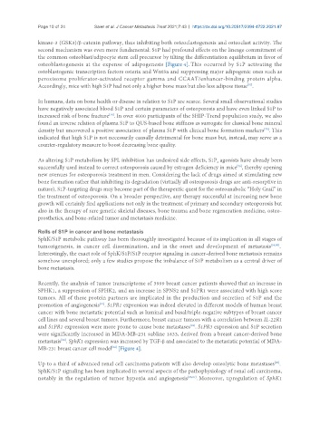
the common osteoblast/adipocyte stem cell precursor by tilting the differentiation equilibrium in favor of
osteoblastogenesis at the expense of adipogenesis [Figure 4]. This occurred by S1P activating the
ostoblastogenic transcription factors osterix and Wnt5a and suppressing major adipogenic ones such as
peroxisome proliferator-activated receptor gamma and CCAAT/enhancer-binding protein alpha.
Accordingly, mice with high S1P had not only a higher bone mass but also less adipose tissue .
[75]
In humans, data on bone health or disease in relation to S1P are scarce. Several small observational studies
have negatively associated blood S1P and certain parameters of osteoporosis and have even linked S1P to
[78]
increased risk of bone fracture . In over 4000 participants of the SHIP-Trend population study, we also
found an inverse relation of plasma S1P to QUS-based bone stiffness as surrogate for classical bone mineral
density but uncovered a positive association of plasma S1P with clinical bone formation markers . This
[75]
indicated that high S1P is not necessarily causally detrimental for bone mass but, instead, may serve as a
counter-regulatory measure to boost decreasing bone quality.
As altering S1P metabolism by SPL inhibition has undesired side effects, S1P agonists have already been
2
successfully used instead to correct osteoporosis caused by estrogen deficiency in mice , thereby opening
[79]
new avenues for osteoporosis treatment in men. Considering the lack of drugs aimed at stimulating new
bone formation rather that inhibiting its degradation (virtually all osteoporosis drugs are anti-resorptive in
nature), S1P-targeting drugs may become part of the therapeutic quest for the osteoanabolic “Holy Grail” in
the treatment of osteoporosis. On a broader perspective, any therapy successful at increasing new bone
growth will certainly find applications not only in the treatment of primary and secondary osteoporosis but
also in the therapy of rare genetic skeletal diseases, bone trauma and bone regeneration medicine, osteo-
prosthetics, and bone-related tumor and metastasis medicine.
Rolls of S1P in cancer and bone metastasis
SphK/S1P metabolic pathway has been thoroughly investigated because of its implication in all stages of
tumorigenesis, in cancer cell dissemination, and in the onset and development of metastasis [56,80] .
Interestingly, the exact role of SphK/S1P/S1P receptor signaling in cancer-derived bone metastasis remains
somehow unexplored; only a few studies propose the imbalance of S1P metabolism as a central driver of
bone metastasis.
Recently, the analysis of tumor transcriptome of 3999 breast cancer patients showed that an increase in
SPHK1, a suppression of SPHK2, and an increase in SPNS2 and S1PR1 were associated with high score
tumors. All of these protein partners are implicated in the production and secretion of S1P and the
[81]
promotion of angiogenesis . S1PR1 expression was indeed elevated in different models of human breast
cancer with bone metastatic potential such as luminal and basal/triple-negative subtypes of breast cancer
cell lines and several breast tumors. Furthermore, breast cancer tumors with a correlation between IL-22R1
and S1PR1 expression were more prone to cause bone metastases . S1PR3 expression and S1P secretion
[82]
were significantly increased in MDA-MB-231 subline 1833, derived from a breast cancer-derived bone
metastasis . SphK1 expression was increased by TGF-β and associated to the metastatic potential of MDA-
[83]
MB-231 breast cancer cell model [Figure 4].
[84]
Up to a third of advanced renal cell carcinoma patients will also develop osteolytic bone metastases .
[85]
SphK/S1P signaling has been implicated in several aspects of the pathophysiology of renal cell carcinoma,
notably in the regulation of tumor hypoxia and angiogenesis [86,87] . Moreover, upregulation of SphK1