
Page 45 - Read Online
P. 45
Saier et al. J Cancer Metastasis Treat 2021;7:43 https://dx.doi.org/10.20517/2394-4722.2021.87 Page 7 of 24
[53]
this class of lipids . Sphingolipid metabolism involves three interconnected pathways, namely the de novo
synthesis pathway, salvage pathway, and sphingomyelinase (SMase) pathway, all of which generate
ceramide from complex lipids that is eventually converted into sphingosine and S1P [54,55] . These pathways
were initially thought to be autonomous of each other in ceramide generation, but the metabolites
synthesized in these three metabolic pathways are highly reversible, non-distinguishable, and hence
interdependent.
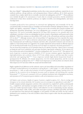
Ceramide produced by these pathways is converted into sphingosine and eventually S1P by the
concatenated effect of enzymes such as ceramidase and functionally redundant sphingosine kinases 1 and 2
(SphK1 and SphK2), respectively [Figure 3]. This process is also reversible and ceramide synthase and
[56]
sphingosine phosphate phosphatase can revert sphingosine into ceramide and S1P to sphingosine,
respectively. S1P can be irreversibly degraded by S1P lyase (SPL) present on the cytosolic side of the
endoplasmic reticulum to form non-sphingolipid products: phospho-ethanolamine and hexadecenal (a fatty
aldehyde). While these enzymes are intracellular, LPPs reside on the cell membrane and keep extracellular
S1P in check [57,58] . Although minimal roles of intracellular S1P signaling have been reported, the major
biological functions of S1P (such as embryonic and postnatal vascular development, vascular integrity and
tone, hematopoiesis and trafficking of immune and stem cells, and platelet formation and activation in
addition to bone homeostasis) are suggested to be receptor dependent and necessitate an efficient export of
S1P into the blood and lymph where the levels of S1P are higher as compared to the tissue parenchyma [58-60] .
The two bona fide transporters of S1P identified thus far are spinster homolog 2 (Spns2) that is expressed
on lymphatic and blood endothelial cells and major facilitator superfamily transporter 2b that exports S1P
from erythrocytes and activated platelets , therefore making these cells “sources of S1P”. This exported S1P
[61]
needs chaperons to promote aqueous solubility of S1P, increase resistance against degradation and
dephosphorylation, and accelerate release of S1P from cellular sources, and they may alter receptor
selectivity and signaling bias [59,62,63] . These chaperons include high-density lipoprotein bound to
apolipoprotein M, albumin, low-density lipoprotein, very low-density lipoprotein, and the recently
identified chaperon apolipoprotein A4 [64,65] . While blood chaperones are well described, less is known about
what chaperones S1P associates within the lymph and interstitial fluids.
Highly regulated local differences in the synthesis, export, and intracellular and extracellular degradation of
S1P lead to marked differences in its abundance among blood (~1 µM), lymph (~0.1 µM), and the tissue
parenchyma (< 1 nM), as well as the formation of local S1P gradients within tissues. Sensing of this S1P
gradient by the receptors drives biological processes such as immune cell trafficking and vascular
homeostasis [58,66,67] . Factors such as presence of de novo synthesis machinery, lack of degradation machinery,
and expression of S1P transporters govern whether a cell can be deemed as a source of S1P.
S1P binds to and activates a family of cognate G-protein coupled receptors, S1P . Due to overlap and
1-5
divergence in Gα subunit selectivity, S1P receptors can act in synergy or in opposition to each other [68,69] .
Notable examples include antagonistic activities of the exclusively Gα-coupled S1P and the predominantly
i
1
Gα -coupled S1P and sometimes synergistic activities of S1P and S1P , which can also couple to Gα. S1P
3
1
1
i
2
12/13
is the most widely expressed receptor, which is well studied for its role in maintaining vascular, immune,
and bone homeostasis. S1PR1, S1PR2, and S1PR3 transcripts in primary osteoblasts and S1PR1 and S1PR2
in osteoclasts have been detected with negligible or null quantity of S1PR4 and S1PR5, but this could be
attributed to poor sensitivity of the tools available to detect these receptors rather than the actual absence of
these receptors.