
Page 86 - Read Online
P. 86
Kurt et al. J Cancer Metastasis Treat 2019;5:8 I http://dx.doi.org/10.20517/2394-4722.2018.80 Page 3 of 6
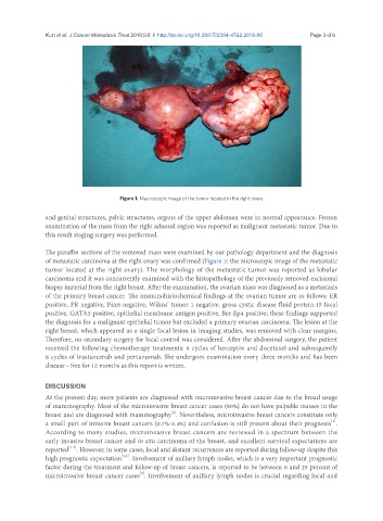
Figure 1. Macroscopic image of the tumor located in the right ovary
and genital structures, pelvic structures, organs of the upper abdomen were in normal appearance. Frozen
examination of the mass from the right adnexal region was reported as malignant metastatic tumor. Due to
this result staging surgery was performed.
The paraffin sections of the removed mass were examined by our pathology department and the diagnosis
of metastatic carcinoma at the right ovary was confirmed (Figure 2: the microscopic image of the metastatic
tumor located at the right ovary). The morphology of the metastatic tumor was reported as lobular
carcinoma and it was concurrently examined with the histopathology of the previously removed excisional
biopsy material from the right breast. After the examination, the ovarian mass was diagnosed as a metastasis
of the primary breast cancer. The immunohistochemical findings at the ovarian tumor are as follows: ER
positive, PR negative, Pax8 negative, Wilms’ tumor 1 negative, gross cystic disease fluid protein-15 focal
positive, GATA3 positive, epithelial membrane antigen positive, Ber-Ep4 positive; these findings supported
the diagnosis for a malignant epithelial tumor but excluded a primary ovarian carcinoma. The lesion at the
right breast, which appeared as a single focal lesion in imaging studies, was removed with clear margins.
Therefore, no secondary surgery for local control was considered. After the abdominal surgery, the patient
received the following chemotherapy treatments: 8 cycles of herceptin and docetaxel and subsequently
8 cycles of trastuzumab and pertuzumab. She undergoes examination every three months and has been
disease - free for 12 months as this report is written.
DISCUSSION
At the present day, more patients are diagnosed with microinvasive breast cancer due to the broad usage
of mammography. Most of the microinvasive breast cancer cases (80%) do not have palpable masses in the
[2]
breast and are diagnosed with mammography . Nevertheless, microinvasive breast cancers constitute only
[1]
a small part of invasive breast cancers (0.7%-2.4%) and confusion is still present about their prognosis .
According to many studies, microinvasive breast cancers are reviewed in a spectrum between the
early invasive breast cancer and in situ carcinoma of the breast, and excellent survival expectations are
[1-4]
reported . However, in some cases, local and distant recurrences are reported during follow-up despite this
[5,6]
high prognostic expectation . Involvement of axillary lymph nodes, which is a very important prognostic
factor during the treatment and follow-up of breast cancers, is reported to be between 0 and 25 percent of
[3]
microinvasive breast cancer cases . Involvement of axillary lymph nodes is crucial regarding local and