
Page 85 - Read Online
P. 85
Page 2 of 6 Kurt et al. J Cancer Metastasis Treat 2019;5:8 I http://dx.doi.org/10.20517/2394-4722.2018.80
[1]
deaths among women . Microinvasive breast cancer makes 0.7%-2.4% of all cases, it is a rare subgroup of
breast cancer. The term of “microinvasive breast cancer” was used by Logios in 1982. Later, the World Health
Organization defined it as an invasive carcinoma of the breast with no invasive focus measuring more than
[2]
1 mm . The prognosis of microinvasive breast cancer is open for discussion, since it is rare and difficult to
diagnose. Generally, it is known that their clinical behavior is between invasive carcinoma and carcinoma
in situ. Lymph node metastasis, which is one of the most important prognostic factors of breast cancer, is
[3]
rare among microinvasive breast cancers (0%-25%) and there is an expectation of better clinical course .
On the other hand, although there are very few in the literature, there are reported cases of aggressively
progressive microinvasive breast cancers that presented clinical findings with lymph node involvement and
[4]
distant organ metastasis .
In this article, we present a case of microinvasive breast cancer that presented clinical findings with
supraclavicular lymph node involvement and ovarian metastasis without any findings from breast
examination. We found it to be interesting, due to the rarity of such cases.
CASE REPORT
A 54-year-old patient (Gravida 4, parity 3, 3 years since menopause, gave birth to her last child 20 years ago)
was visited due to the complaint of a palpable lymph node at the right supraclavicular region. Due to the
finding of a metastatic carcinoma at the lymph node after thin needle aspiration biopsy, she was investigated
with imaging and systemic examination in order to find the primary focus. Systemic examination only
demonstrated a palpable lymph node, which was 2 cm × 3 cm in size, at the right supraclavicular region. No
other abnormal clinical finding was detected. A solid, mobile mass was found at the right adnexal region
during gynecologic examination. The mass was 6 cm × 7 cm in size. Pelvic ultrasonography of the left
adnexa and the uterus was normal regarding the patient’s age. The lesion, which seemed to be originating
from the right ovary was measured to be 65 mm × 72 mm in size. No free abdominal fluid was present.
Ultrasonography of the upper abdomen and the physical examination of the breast was normal. Cervical
smear, endometrial sampling, endoscopy of the gastrointestinal system, routine biochemical tests and
complete blood count resulted in the normal range.
Apart from elevated CA15-3 concentration (157 IU/mL) and erythrocyte sedimentation rate (72 mm per
30 min); no abnormal blood parameters were present. Bilateral mammography was reviewed as Breast
Imaging Reporting and Data System - 4B for the right breast (possible malignant findings), and therefore
excisional biopsy (surgical margin clear of disease) was performed. Following radiological examinations
showed no residual lesions. Pathological examination resulted as: microinvasive breast carcinoma, nuclear
grade II. Immunohistochemistry results were as follows: p63 (+) at myoepithelial cells, estrogen receptor (ER)
(+) for about 5%-10% of the cells at the ductal carcinoma in situ (DCIS) focus, progesterone receptor (PR) (-)
for the cells at the DCIS focus.
Positron emission tomography-computerized tomography (PET-CT) showed increased F-18 fluorodeoxyglucose
(FDG) uptake at both supraclavicular lymph nodes, especially more prominent on the right cervical level. In
addition, increased uptake of FDG was also present at the lymph nodes in thorax and abdomen. The mass
lesion, whose margins were not distinguishable from the uterus, was located in the right adnexal region and
showed increased F-18 FDG uptake. These radiological findings in the right adnexal region supported the
diagnosis of primary gynecological malignancy.
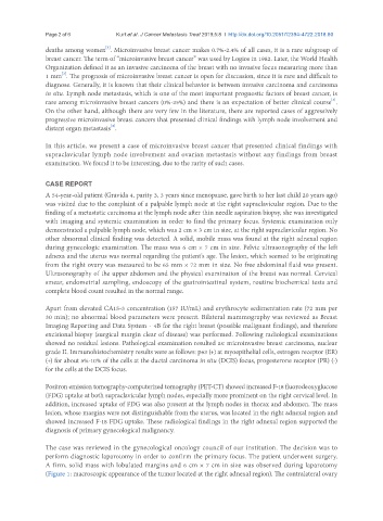
The case was reviewed in the gynecological oncology council of our institution. The decision was to
perform diagnostic laparotomy in order to confirm the primary focus. The patient underwent surgery.
A firm, solid mass with lobulated margins and 6 cm × 7 cm in size was observed during laparotomy
(Figure 1: macroscopic appearance of the tumor located at the right adnexal region). The contralateral ovary