
Page 62 - Read Online
P. 62
Anand et al. J Cancer Metastasis Treat 2019;5:6 I http://dx.doi.org/10.20517/2394-4722.2018.98 Page 7 of 14
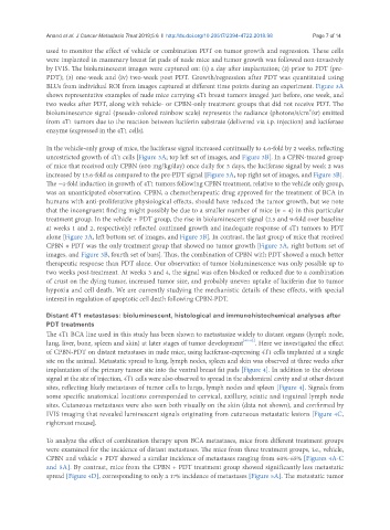
used to monitor the effect of vehicle or combination PDT on tumor growth and regression. These cells
were implanted in mammary breast fat pads of nude mice and tumor growth was followed non-invasively
by IVIS. The bioluminescent images were captured on: (1) a day after implantation; (2) prior to PDT (pre-
PDT); (3) one-week and (iv) two-week post PDT. Growth/regression after PDT was quantitated using
BLUs from individual ROI from images captured at different time points during an experiment. Figure 3A
shows representative examples of nude mice carrying 4T1 breast tumors imaged just before, one week, and
two weeks after PDT, along with vehicle- or CPBN-only treatment groups that did not receive PDT. The
2
bioluminescence signal (pseudo-colored rainbow scale) represents the radiance (photons/s/cm /sr) emitted
from 4T1 tumors due to the reaction between luciferin substrate (delivered via i.p. injection) and luciferase
enzyme (expressed in the 4T1 cells).
In the vehicle-only group of mice, the luciferase signal increased continually to 4.6-fold by 2 weeks, reflecting
unrestricted growth of 4T1 cells [Figure 3A; top left set of images, and Figure 3B]. In a CPBN-treated group
of mice that received only CPBN (600 mg/kg/day) once daily for 3 days, the luciferase signal by week 2 was
increased by 13.6-fold as compared to the pre-PDT signal [Figure 3A, top right set of images, and Figure 3B].
The ~4-fold induction in growth of 4T1 tumors following CPBN treatment, relative to the vehicle only group,
was an unanticipated observation. CPBN, a chemotherapeutic drug approved for the treatment of BCA in
humans with anti-proliferative physiological effects, should have reduced the tumor growth, but we note
that the incongruent finding might possibly be due to a smaller number of mice (n = 4) in this particular
treatment group. In the vehicle + PDT group, the rise in bioluminescent signal (2.5 and 9-fold over baseline
at weeks 1 and 2, respectively) reflected continued growth and inadequate response of 4T1 tumors to PDT
alone [Figure 3A, left bottom set of images, and Figure 3B]. In contrast, the last group of mice that received
CPBN + PDT was the only treatment group that showed no tumor growth [Figure 3A, right bottom set of
images, and Figure 3B, fourth set of bars]. Thus, the combination of CPBN with PDT showed a much better
therapeutic response than PDT alone. Our observation of tumor bioluminescence was only possible up to
two weeks post-treatment. At weeks 3 and 4, the signal was often blocked or reduced due to a combination
of crust on the dying tumor, increased tumor size, and probably uneven uptake of luciferin due to tumor
hypoxia and cell death. We are currently studying the mechanistic details of these effects, with special
interest in regulation of apoptotic cell death following CPBN-PDT.
Distant 4T1 metastases: bioluminescent, histological and immunohistochemical analyses after
PDT treatments
The 4T1 BCA line used in this study has been shown to metastasize widely to distant organs (lymph node,
lung, liver, bone, spleen and skin) at later stages of tumor development [40-42] . Here we investigated the effect
of CPBN-PDT on distant metastases in nude mice, using luciferase-expressing 4T1 cells implanted at a single
site on the animal. Metastatic spread to lung, lymph nodes, spleen and skin was observed at three weeks after
implantation of the primary tumor site into the ventral breast fat pads [Figure 4]. In addition to the obvious
signal at the site of injection, 4T1 cells were also observed to spread in the abdominal cavity and at other distant
sites, reflecting likely metastases of tumor cells to lungs, lymph nodes and spleen [Figure 4]. Signals from
some specific anatomical locations corresponded to cervical, axillary, sciatic and inguinal lymph node
sites. Cutaneous metastases were also seen both visually on the skin (data not shown), and confirmed by
IVIS imaging that revealed luminescent signals originating from cutaneous metastatic lesions [Figure 4C,
rightmost mouse].
To analyze the effect of combination therapy upon BCA metastases, mice from different treatment groups
were examined for the incidence of distant metastases. The mice from three treatment groups, i.e., vehicle,
CPBN and vehicle + PDT showed a similar incidence of metastases ranging from 60%-65% [Figures 4A-C
and 5A]. By contrast, mice from the CPBN + PDT treatment group showed significantly less metastatic
spread [Figure 4D], corresponding to only a 17% incidence of metastases [Figure 5A]. The metastatic tumor