
Page 66 - Read Online
P. 66
Anand et al. J Cancer Metastasis Treat 2019;5:6 I http://dx.doi.org/10.20517/2394-4722.2018.98 Page 11 of 14
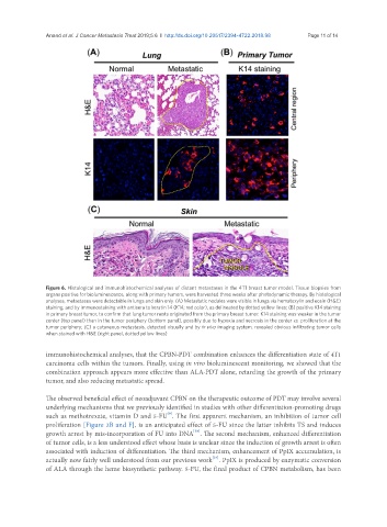
Figure 6. Histological and immunohistochemical analyses of distant metastases in the 4T1 breast tumor model. Tissue biopsies from
organs positive for bioluminescence, along with primary tumors, were harvested three weeks after photodynamic therapy. By histological
analyses, metastases were detectable in lungs and skin only. (A) Metastatic nodules were visible in lungs via hematoxylin and eosin (H&E)
staining, and by immunostaining with antisera to keratin 14 (K14; red color), as delineated by dotted yellow lines; (B) positive K14 staining
in primary breast tumor, to confirm that lung tumor nests originated from the primary breast tumor. K14 staining was weaker in the tumor
center (top panel) than in the tumor periphery (bottom panel), possibly due to hypoxia and necrosis in the center vs. proliferation at the
tumor periphery; (C) a cutaneous metastasis, detected visually and by in vivo imaging system, revealed obvious infiltrating tumor cells
when stained with H&E (right panel, dotted yellow lines)
immunohistochemical analyses, that the CPBN-PDT combination enhances the differentiation state of 4T1
carcinoma cells within the tumors. Finally, using in vivo bioluminescent monitoring, we showed that the
combination approach appears more effective than ALA-PDT alone, retarding the growth of the primary
tumor, and also reducing metastatic spread.
The observed beneficial effect of neoadjuvant CPBN on the therapeutic outcome of PDT may involve several
underlying mechanisms that we previously identified in studies with other differentiation-promoting drugs
[9]
such as methotrexate, vitamin D and 5-FU . The first apparent mechanism, an inhibition of tumor cell
proliferation [Figure 2B and F], is an anticipated effect of 5-FU since the latter inhibits TS and induces
[38]
growth arrest by mis-incorporation of FU into DNA . The second mechanism, enhanced differentiation
of tumor cells, is a less understood effect whose basis is unclear since the induction of growth arrest is often
associated with induction of differentiation. The third mechanism, enhancement of PpIX accumulation, is
[24]
actually now fairly well understood from our previous work . PpIX is produced by enzymatic conversion
of ALA through the heme biosynthetic pathway. 5-FU, the final product of CPBN metabolism, has been