
Page 60 - Read Online
P. 60
Anand et al. J Cancer Metastasis Treat 2019;5:6 I http://dx.doi.org/10.20517/2394-4722.2018.98 Page 5 of 14
(A) (B) (C) (D)
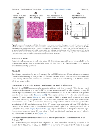
Figure 1. Analyses of protoporphyrin IX (PpIX) in murine breast tumor model. (A) Photograph of a nude female mouse with 4T1
subcutaneous tumors in breast fat pad; (B) hematoxylin and eosin (H&E) stained 4T1 tumor section showing histological details of the
murine breast carcinoma shown in (A); (C) confocal micrographs of frozen sections of 4T1 tumors after oral treatment with vehicle (top)
or capecitabine (CPBN) (bottom) followed by aminolevulinic acid; red signal is mitochondrial PpIX; (D) quantitation of PpIX-specific
fluorescence from confocal micrographs shown in (C); integrated fluorescence intensity was measured using IPLab software. Mean SEM
(18 images from 6 tumors) from three independent experiments is shown. *P = 0.0001
Statistical analyses
Statistical analyses were performed using a two-sided t-test to compare differences between PpIX levels,
expression of markers by immunohistochemistry, cell death and tumor bioluminescence. P ≤ 0.05 was
considered statistically significant.
RESULTS
Experiments were designed to test our hypothesis that oral CPBN, given as a differentiation-promoting agent
(instead of administering its final product 5-FU{Anand, 2017 #100;Maytin, 2018 #114}), may enhance the PS
levels, subsequent cell death response and will impact distant metastases following ALA-PDT in a murine
4T1 breast tumor model.
Combination of oral CPBN before ALA enhances PpIX levels in 4T1 tumors
To study if oral CPBN can successfully replace its relatively toxic final product 5-FU for the purpose of
promoting differentiation prior to ALA-PDT, a murine breast tumor cell line (4T1) equivalent to stage IV
human triple negative breast carcinoma was injected in the breast fad pad of female nude mice to generate
a murine breast tumor model [Figure 1A and B]. Following the injection of 4T1 cells, mice with visible and
palpable 4T1 breast tumors [Figure 1A] were given either CPBN or vehicle (orally by gavage, once a day
for 3 days), followed by intraperitoneal ALA administration on the 4th day. Tumors were harvested and
frozen sections were analyzed by confocal microscopy using excitation and emission settings that allow
visualization of PpIX-specific fluorescence. In the 4T1 tumors from mice treated with oral CPBN, PpIX was
more abundant and also present throughout the tumor at much higher levels, as compared with vehicle
treated (control) tumors [Figure 1C; bottom vs. Top]. Digital quantification of the PpIX fluorescence signal
from confocal images confirmed this observation by showing a statistically significant increase of ~4-fold in
PpIX levels in CPBN treated tumors [Figure 1D].
CPBN pretreatment enhances differentiation, inhibits proliferation and enhances cell death
following PDT
5FU, a chemotherapeutic drug and the final product of CPBN metabolism specifically converted in the
tumors due to high levels of CDA, and TYMP [28,30] , is known to downregulate DNA synthesis through