
Page 43 - Read Online
P. 43
Tu et al. J Cancer Metastasis Treat 2018;4:58 I http://dx.doi.org/10.20517/2394-4722.2018.67 Page 9 of 16
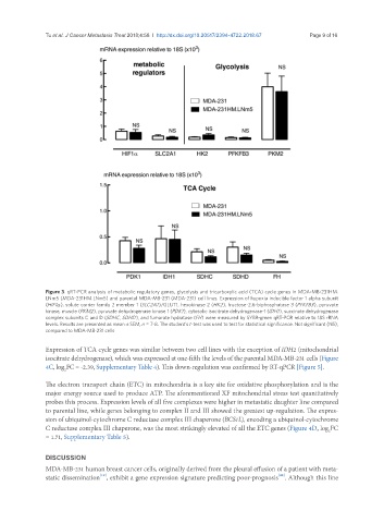
Figure 3. qRT-PCR analysis of metabolic regulatory genes, glycolysis and tricarboxylic acid (TCA) cycle genes in MDA-MB-231HM.
LNm5 (MDA-231HM.LNm5) and parental MDA-MB-231 (MDA-231) cell lines. Expression of hypoxia inducible factor 1 alpha subunit
(HIF1a ), solute carrier family 2 member 1 (SLC2A1)/GLUT1, hexokinase 2 (HK2), fructose-2,6-biphosphatase 3 (PFKFB3), pyruvate
kinase, muscle (PKM2), pyruvate dehydrogenase kinase 1 (PDK1), cytosolic isocitrate dehydrogenase-1 (IDH1), succinate dehydrogenase
complex subunits C and D (SDHC, SDHD), and fumarate hydratase (FH) were measured by SYBR-green qRT-PCR relative to 18S rRNA
levels. Results are presented as mean ± SEM, n = 7-8. The student’s t-test was used to test for statistical significance. Not significant (NS),
compared to MDA-MB-231 cells
Expression of TCA cycle genes was similar between two cell lines with the exception of IDH2 (mitochondrial
isocitrate dehydrogenase), which was expressed at one-fifth the levels of the parental MDA-MB-231 cells (Figure
4C, log FC = -2.39, Supplementary Table 4). This down-regulation was confirmed by RT-qPCR [Figure 5].
2
The electron transport chain (ETC) in mitochondria is a key site for oxidative phosphorylation and is the
major energy source used to produce ATP. The aforementioned XF mitochondrial stress test quantitatively
probes this process. Expression levels of all five complexes were higher in metastatic daughter line compared
to parental line, while genes belonging to complex II and III showed the greatest up-regulation. The expres-
sion of ubiquinol-cytochrome C reductase complex III chaperone (BCS1L), encoding a ubiquinol-cytochrome
C reductase complex III chaperone, was the most strikingly elevated of all the ETC genes (Figure 4D, log FC
2
= 1.71, Supplementary Table 5).
DISCUSSION
MDA-MB-231 human breast cancer cells, originally derived from the pleural effusion of a patient with meta-
[40]
[19]
static dissemination , exhibit a gene expression signature predicting poor-prognosis . Although this line