
Page 502 - Read Online
P. 502
Zeng et al. Hepatoma Res2020;6:43 I http://dx.doi.org/10.20517/2394-5079.2020.29 Page 9 of 12
A B C
D E F
G H I
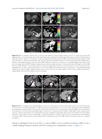
Figure 10. SBRT for repeated de novo HCC in a 63-year-old man with previous surgical resection of HCC. A: Axial arterial phase MRI
image showed a 1.2-cm enhancing nodule in the right hepatic lobe, adjacent to the diaphragm; B: the patient received his first course of
SBRT with a dose of 48 Gy in six fractions; C: axial arterial phase MRI image 4 years after SBRT demonstrated complete response of this
right lobe lesion; D: axial arterial phase MRI image two years after the first SBRT showed a 1-cm enhancing nodule in the caudate lobe of
the liver; E: the patient received a second course of SBRT with a dose of 48 Gy in six fractions; F: axial arterial phase MRI image 3 years
after the second course of SBRT revealed complete response of the caudate lobe lesion; G: axial arterial phase MRI image 18 months after
the second SBRT showed a 1.2-cm enhancing lesion in the right hepatic lobe, as well as an additional lesion at a lower level (not shown),
suggesting de novo HCC; H: the patient received a third course of SBRT with a dose of 54 Gy in nine fractions. I: axial arterial phase MRI
image one year after this SBRT revealed complete tumor response of the de novo lesion. SBRT: stereotactic body radiation therapy; HCC:
hepatocellular carcinoma; MRI: magnetic resonance imaging
A B C
D E F
Figure 11. SBRT as a bridge to liver transplantation for HCC in a 55-year-old man with alcohol-related cirrhosis and Child-Pugh Class
C liver function. A: Axial arterial phase CT image showed hyper enhancement of a 2 cm mass in the right hepatic lobe (arrowhead),
accompanied by moderate ascites. Liver function was scored as Child-Pugh Class C; B: the patient underwent SBRT with a dose of 50 Gy
in five fractions; C-E: Axial arterial phase MRI images 1.5 months (C), 5 months (D), and 9 months (E) after SBRT showed complete tumor
response. However, focal reaction with enhancement indicated congestion of the hepatic parenchyma in the radiation field. The patient
received a liver transplant 22 months after SBRT; F: axial arterial phase CT image one year after liver transplantation demonstrated a
normal liver. SBRT: stereotactic body radiation therapy; HCC: hepatocellular carcinoma; MRI: magnetic resonance imaging; CT: computed
tomography
difficult to distinguish from de novo HCC, a course of SBRT can be considered. In addition, SBRT can be a
suitable bridging therapy for patients with HCC awaiting liver transplantation [Case 11; Figure 11].