
Page 499 - Read Online
P. 499
Page 6 of 12 Zeng et al. Hepatoma Res2020;6:43 I http://dx.doi.org/10.20517/2394-5079.2020.29
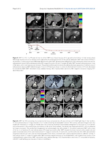
A B
C D E
F
Figure 4. SBRT for HCC in a 99-year-old man for whom SBRT was chosen because of his age and comorbidities. A: Axial venous phase
MRI image showed a 6-cm low-density lesion adjacent to the diaphragm (arrow); B: the patient underwent SBRT with a dose of 54 Gy in
six fractions; C: axial arterial phase MRI image three months after SBRT demonstrated reduced size of the intrahepatic lesion and vascular
enhancement (arrow); D,E: axial arterial phase MRI images 10 months (D) and 17 months (E) after SBRT showed further reduction in size
of the tumor, which was hypovascular (arrows). The perilesional liver parenchyma in the enhanced phase appeared as ill-defined areas of
enhancement, favoring SBRT-related changes; F: Serum AFP levels are shown in relation to the treatment timeline. AFP levels decreased
to normal after SBRT. SBRT: stereotactic body radiation therapy; HCC: hepatocellular carcinoma; MRI: magnetic resonance imaging; AFP:
alpha-fetoprotein
A B
C D E
F G H
Figure 5. SBRT for HCC consisting of an extensive fat-tissue component in a 41-year-old man with Child-Pugh Class C liver function.
A: Axial arterial phase CT image showed a 2-cm nodule containing fat attenuation (arrow). Mild ascites and splenomegaly were also
evident; B: multiphasic CT images six months after liver protecting treatment demonstrated enlargement of the lesion to 3 cm (arrow),
with enhancement in the arterial phase and washout in the portal venous phase. The patient was diagnosed with HCC with an extensive
fat-tissue component. The ascites had disappeared, but splenomegaly was still present; C: the patient underwent SBRT with a dose
of 54 Gy in six fractions; D: axial arterial phase CT image two months after SBRT showed the lesion (arrow) with a stable size and
partial devascularization; E-G: axial arterial phase CT images 6 months (E), 9 months (F), and 13 months (G) after SBRT demonstrated
progressive reduction in tumor size and devascularization of the lesion, consistent with a partial response based on mRECIST criteria; H:
axial arterial phase CT image 25 months after SBRT showed complete response of the treated lesion, with marked focal atrophy of the
irradiated hepatic parenchyma. SBRT: stereotactic body radiation therapy; HCC: hepatocellular carcinoma; CT: computed tomography;
mRECIST: modified Response Evaluation Criteria In Solid Tumor