
Page 497 - Read Online
P. 497
Page 4 of 12 Zeng et al. Hepatoma Res2020;6:43 I http://dx.doi.org/10.20517/2394-5079.2020.29
A B C
D E F
G H I
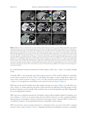
Figure 1. SBRT for HCC in a 59-year-old man who refused surgical resection and RFA. A: Axial arterial phase MRI image showed
hyperenhancement of a 1.8-cm liver nodule in the right lobe of the liver; B: the patient received SBRT with a dose of 50 Gy in five fractions; C:
axial arterial phase MRI image 2 months after SBRT revealed a stable-size, enhancing lesion (arrowhead). The normal liver parenchyma
received 25 Gy and appeared as a low-density area, indicating acute radiation injury; D: axial arterial phase MRI image 4 months after
SBRT showed decreased size of the tumor, with atrophy of the perilesional hepatic parenchyma; E: axial arterial phase MRI image
6 months after SBRT showed necrotic changes and a hypovascular target lesion. The perilesional hepatic parenchyma, which received
high-dose radiation, also exhibited necrotic changes; F: axial arterial phase MRI image nine months after SBRT demonstrated complete
regression of the treated lesion and atrophy of the irradiated hepatic parenchyma; G: axial arterial phase CT image 18 months after SBRT
showed a scar in the radiation field. Complete repair of the radiation damage was also observed; H,I: axial arterial phase MRI images
6 years (H) and 7 years (I) after SBRT demonstrated a stable lesion size but the presence of complete necrosis and ring enhancement,
indicating a complete radiologic response. Note the SBRT-related changes, which can be differentiated from tumor recurrence. SBRT:
stereotactic body radiation therapy; HCC: hepatocellular carcinoma; RFA: radiofrequency ablation; MRI: magnetic resonance imaging; CT:
computed tomography
be a useful alternative treatment in patients who refuse surgery or RFA. Case 1 [Figure 1] is a typical example
of this.
Currently, SBRT is most frequently used when surgical resection or RFA would be difficult or unfeasible,
as with tumors located at the center of liver, in the hepatic hilar region, or close to large blood vessels (as in
Cases 2 and 3, which are shown in Figures 2 and 3). It is also commonly used in patients who are older [Case
4; Figure 4] or have significant comorbidities, such as poor liver function [Case 5; Figure 5].
SBRT may also be used for residual cancer after surgical resection, as in Case 6 [Figure 6], or after RFA, as in
Case 7 [Figure 7]. In these situations, the tumor is often expected to be difficult to treat with surgery or RFA,
but these treatments are tried initially. When residual tumor is found during follow-up, SBRT will generally
be the most appropriate treatment.
SBRT may serve as adjuvant treatment for intrahepatic tumors with incomplete iodized oil retention, as in
Case 8 [Figure 8]. TACE can be used to reduce the size of HCCs; however, it is often considered palliative
therapy because of incomplete iodized oil retention by intrahepatic tumors. Adjuvant SBRT can function as
consolidation treatment, converting palliative therapy to potentially curative therapy.
SBRT has also been used as salvage treatment for intrahepatic tumor recurrence after RFA or surgical
resection [Cases 9 and 10; Figures 9 and 10]. Although metachronous intrahepatic recurrence is sometimes