
Page 501 - Read Online
P. 501
Page 8 of 12 Zeng et al. Hepatoma Res2020;6:43 I http://dx.doi.org/10.20517/2394-5079.2020.29
A B C
D E F
G H
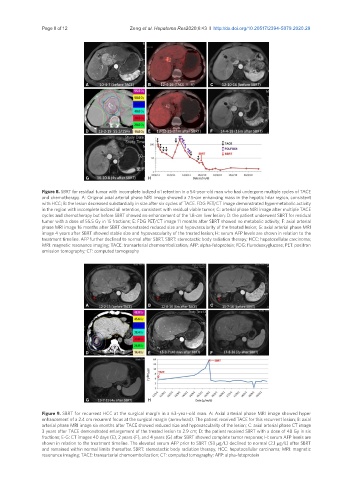
Figure 8. SBRT for residual tumor with incomplete iodized oil retention in a 54-year-old man who had undergone multiple cycles of TACE
and chemotherapy. A: Original axial arterial phase MRI image showed a 7.5-cm enhancing mass in the hepatic hilar region, consistent
with HCC; B: the lesion decreased substantially in size after six cycles of TACE. FDG PET/CT image demonstrated hypermetabolic activity
in the region with incomplete iodized oil retention, consistent with residual viable tumor; C: arterial phase MRI image after multiple TACE
cycles and chemotherapy but before SBRT showed no enhancement of the 1.8-cm liver lesion; D: the patient underwent SBRT for residual
tumor with a dose of 55.5 Gy in 15 fractions; E: FDG PET/CT image 11 months after SBRT showed no metabolic activity; F: axial arterial
phase MRI image 16 months after SBRT demonstrated reduced size and hypovascularity of the treated lesion; G: axial arterial phase MRI
image 4 years after SBRT showed stable size and hypovascularity of the treated lesion; H: serum AFP levels are shown in relation to the
treatment timeline. AFP further declined to normal after SBRT. SBRT: stereotactic body radiation therapy; HCC: hepatocellular carcinoma;
MRI: magnetic resonance imaging; TACE: transarterial chemoembolization; AFP: alpha-fetoprotein; FDG: flurodeoxyglucose; PET: positron
emission tomography; CT: computed tomography
A B C
D E F
G H
Figure 9. SBRT for recurrent HCC at the surgical margin in a 63-year-old man. A: Axial arterial phase MRI image showed hyper
enhancement of a 2.4 cm recurrent focus at the surgical margin (arrowhead). The patient received TACE for this recurrent lesion; B: axial
arterial phase MRI image six months after TACE showed reduced size and hypovascularity of the lesion; C: axial arterial phase CT image
3 years after TACE demonstrated enlargement of the treated lesion to 2.9 cm; D: the patient received SBRT with a dose of 48 Gy in six
fractions; E-G: CT images 40 days (E), 2 years (F), and 4 years (G) after SBRT showed complete tumor response; H: serum AFP levels are
shown in relation to the treatment timeline. The elevated serum AFP prior to SBRT (53 µg/L) declined to normal (2.1 µg/L) after SBRT
and remained within normal limits thereafter. SBRT: stereotactic body radiation therapy; HCC: hepatocellular carcinoma; MRI: magnetic
resonance imaging; TACE: transarterial chemoembolization; CT: computed tomography; AFP: alpha-fetoprotein