
Page 498 - Read Online
P. 498
Zeng et al. Hepatoma Res2020;6:43 I http://dx.doi.org/10.20517/2394-5079.2020.29 Page 5 of 12
A B C
D E F
G
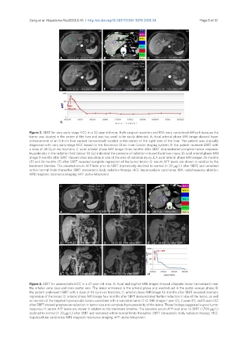
Figure 2. SBRT for very early-stage HCC in a 52-year-old man. Both surgical resection and RFA were considered difficult because the
tumor was located in the center of the liver and was too small to be easily detected. A: Axial arterial phase MRI image showed hyper
enhancement of an 0.8-cm liver nodule (arrowhead) located in the center of the right lobe of the liver. The patient was clinically
diagnosed with very early-stage HCC based on the Barcelona Clinic Liver Cancer staging system; B: the patient received SBRT with
a dose of 48 Gy in six fractions; C: axial arterial phase MRI image three months after SBRT demonstrated complete tumor response.
Hypodensity in the radiation field (about 30 Gy) indicated the presence of radiation-induced focal liver injury; D: axial arterial phase MRI
image 9 months after SBRT showed clear reduction in size of the area of radiation injury; E,F: axial arterial phase MRI images 26 months
(E) and 56 months (F) after SBRT revealed complete regression of the tumor lesion; G: serum AFP levels are shown in relation to the
treatment timeline. The elevated serum AFP level prior to SBRT dramatically declined to normal (< 20 µg/L) after SBRT, and remained
within normal limits thereafter. SBRT: stereotactic body radiation therapy; HCC: hepatocellular carcinoma; RFA: radiofrequency ablation;
MRI: magnetic resonance imaging; AFP: alpha-fetoprotein
A B
C D E
F G H
Figure 3. SBRT for unresectable HCC in a 47-year-old man. A: Axial and sagittal MRI images showed a hepatic lesion (arrowhead) near
the inferior vena cava and main portal vein. The lesion enhanced in the arterial phase and washed out in the portal venous phase; B:
the patient underwent SBRT with a dose of 45 Gy in six fractions; C: arterial phase MRI image 1.5 months after SBRT revealed dramatic
regression of the lesion; D: arterial phase MRI image four months after SBRT demonstrated further reduction in size of the lesion, as well
as necrosis of the targeted hypovascular lesion, consistent with a nonviable tumor; E-G: MRI images 1 year (E), 2 years (F), and 3 years (G)
after SBRT showed progressive reduction in tumor size and complete hypovascularity of the lesion. These findings suggested a good tumor
response; H: serum AFP levels are shown in relation to the treatment timeline. The elevated serum AFP level prior to SBRT (1,709 µg/L)
declined to normal (< 20 µg/L) after SBRT and remained within normal limits thereafter. SBRT: stereotactic body radiation therapy; HCC:
hepatocellular carcinoma; MRI: magnetic resonance imaging; AFP: alpha-fetoprotein