
Page 507 - Read Online
P. 507
Page 2 of 8 Yip et al. Hepatoma Res 2020;6:44 I http://dx.doi.org/10.20517/2394-5079.2020.30
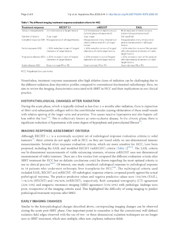
Table 1. The different imaging treatment response evaluation criteria for HCC
Treatment response RECIST 1.1 mRECIST EASL
Tumour measurements Uni-dimensional of target lesions Uni-dimensional of viable tumours Bi-dimensional of viable tumours
(arterial phase enhancement) (arterial phase enhancement)
Number of lesions 2 per organ 2 per organ Not specified
Complete response (CR) Disappearance of all target lesions Disappearance of any intratumoral Disappearance of any intratumoral
arterial enhancement in all target arterial enhancement in all target
lesions lesions
Partial response (PR) ≥ 30% reduction in sum of longest ≥ 30% reduction in sum of longest ≥ 50% reduction in sum of the product
diameters of target lesions diameters of viable target lesions of bi-dimensional diameters of viable
target lesions
Progressive disease (PD) ≥ 20% increase in sum of longest ≥ 20% increase in sum of longest ≥ 25% increase in sum of the product
diameters of target lesions diameters of viable target lesions of bi-dimensional diameters of viable
target lesions
Stable disease (SD) Does not meet PR or PD Does not meet PR or PD Does not meet PR or PD
HCC: Hepatocellular carcinoma
Nonetheless, treatment response assessment after high ablative doses of radiation can be challenging due to
the different radiation dose deposition profiles compared to conventional fractionated radiotherapy. Here, we
aim to review the imaging characteristics associated with SBRT in HCC and their implications in our clinical
practice.
HISTOPATHOLOGICAL CHANGES AFTER RADIATION
During the acute phase, which is typically defined as less than 3-4 months after radiation, there is deposition
of fibrin and subsequently collagen within the centrilobular venules causing obliteration of these small vessels
with relative sparing of the larger veins and arterioles. This causes reactive hyperaemia and also hepatic cell
[7,8]
loss within the liver . This is collectively known as veno-occlusive disease. In the chronic phase, there is
[8]
significant reduction of hyperaemia with some degree of hyperplasia and parenchymal fibrosis .
IMAGING RESPONSE ASSESSMENT CRITERIA
Although RECIST 1.1 is a universally accepted set of radiological response evaluation criteria in solid
[9]
tumours , these criteria do not apply well in HCC as they are based solely on uni-dimensional tumour
measurements. Several other response evaluation criteria, which are more sensitive for HCC, have been
proposed, including the EASL and modified RECIST (mRECIST) criteria [Table 1] [10,11] . The EASL criteria
use bi-dimensional measurements of viable enhancing tumours, whereas mRECIST uses uni-dimensional
measurement of viable tumours. There are a few studies that compared the different evaluation criteria after
SBRT treatment for HCC but no definite conclusions could be drawn regarding the most optimal criteria to
use in clinical practice [12,13] . Of interest, one study correlated radiological response to pathological response
[14]
in 38 patients who underwent orthotopic liver transplants for HCC . The radiological criteria used
included EASL, RECIST and mRECIST. All radiological response criteria compared poorly against the actual
pathological response. The positive predictive values and negative predictive values were 73%/29% (EASL),
71%/32% (RECIST) and 74%/40% (mRECIST), respectively. Both computed tomography (CT) agreement
(22%-39%) and magnetic resonance imaging (MRI) agreement (31%-39%) with pathologic findings were
poor, irrespective of the imaging criteria used. This highlighted the difficulty of using imaging to predict
pathological treatment response after SBRT.
EARLY IMAGING CHANGES
Similar to the histopathological changes described above, corresponding imaging changes can be observed
during the acute post-SBRT phase. One important point to remember is that the conventional well-defined
radiation field edges observed with the use of two- or three-dimensional radiation techniques are no longer
seen in SBRT treatment, which uses multiple, often non-coplanar, radiation fields.