
Page 510 - Read Online
P. 510
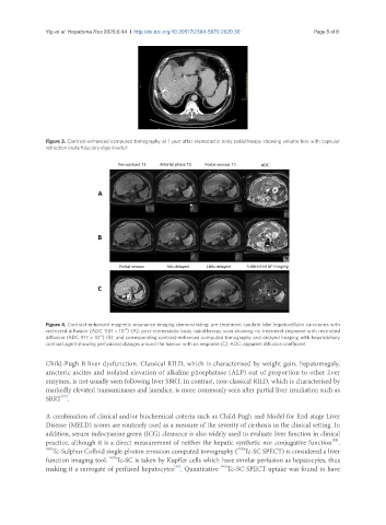
Yip et al. Hepatoma Res 2020;6:44  I  http://dx.doi.org/10.20517/2394-5079.2020.30                                                    Page 5 of 8


               Figure 3. Contrast-enhanced computed tomography at 1 year after stereotactic body radiotherapy showing volume loss with capsular
               retraction (note fiduciary clips in-situ)


               Figure 4. Contrast-enhanced magnetic resonance imaging demonstrating: pre-treatment caudate lobe hepatocellular carcinoma with
                                       -6
               restricted diffusion (ADC 1101 × 10 ) (A); post-stereotactic body radiotherapy scan showing no treatment response with restricted
                                -6
               diffusion (ADC 917 × 10 ) (B); and corresponding contrast-enhanced computed tomography and delayed imaging with hepatobiliary
               contrast agent showing perfusional changes around the tumour with no response (C). ADC: apparent diffusion coefficient
               Child-Pugh B liver dysfunction. Classical RILD, which is characterised by weight gain, hepatomegaly,
               anicteric ascites and isolated elevation of alkaline phosphatase (ALP) out of proportion to other liver
               enzymes, is not usually seen following liver SBRT. In contrast, non-classical RILD, which is characterised by
               markedly elevated transaminases and jaundice, is more commonly seen after partial liver irradiation such as
                    [27]
               SBRT .
               A combination of clinical and/or biochemical criteria such as Child-Pugh and Model for End-stage Liver
               Disease (MELD) scores are routinely used as a measure of the severity of cirrhosis in the clinical setting. In
               addition, serum indocyanine green (ICG) clearance is also widely used to evaluate liver function in clinical
               practice, although it is a direct measurement of neither the hepatic synthetic nor conjugative function .
                                                                                                       [28]
               99m Tc-Sulphur Colloid single-photon emission computed tomography ( Tc-SC SPECT) is considered a liver
                                                                          99m
               function imaging tool.  Tc-SC is taken by Kupffer cells which have similar perfusion as hepatocytes, thus
                                   99m
                                                                     99m
                                                       [29]
               making it a surrogate of perfused hepatocytes . Quantitative  Tc-SC SPECT uptake was found to have