
Page 277 - Read Online
P. 277
Magee et al. Egr1 in liver metabolism and cancer
S350 T391
T309 S378
T526
Amino acid 1 281 314 543
K272
338 362 390 396 418
Casein kinase II phosphorylation 368
AKT phosphorylation
SUMOylation
Repressor domain
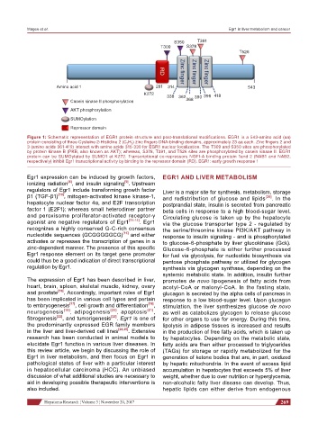
Figure 1: Schematic representation of EGR1 protein structure and post-translational modifications. EGR1 is a 543-amino acid (aa)
protein consisting of three Cysteine 2-Histidine 2 (C 2 H 2 ) zinc fingers DNA-binding domains, approximately 23 aa each. Zinc fingers 2 and
3 (amino acids 361-419) interact with amino acids 315-330 for EGR1 nuclear localization. The T309 and S350 sites are phosphorylated
by protein kinase B (PKB, also known as AKT); whereas, S378, T391, and T526 sites are phosphorylated by casein kinase II. EGR1
protein can be SUMOylated by SUMO1 at K272. Transcriptional co-repressors NGFI-A binding protein 1and 2 (NAB1 and NAB2,
respectively) inhibit Egr1 transcriptional activity by binding to the repressor domain (RD). EGR1: early growth response 1
Egr1 expression can be induced by growth factors, EGR1 AND LIVER METABOLISM
[8]
ionizing radiation , and insulin signaling . Upstream
[9]
regulators of Egr1 include transforming growth factor Liver is a major site for synthesis, metabolism, storage
β1 (TGF-β1) [10] , mitogen-activated kinase kinase-1, and redistribution of glucose and lipids [26] . In the
hepatocyte nuclear factor 4α, and E2F transcription postprandial state, insulin is secreted from pancreatic
factor 1 (E2F1); whereas small heterodimer partner beta cells in response to a high blood-sugar level.
and peroxisome proliferator-activated receptor-γ Circulating glucose is taken up by the hepatocyte
agonist are negative regulators of Egr1 [11-14] . Egr1 via the glucose transporter type 2 - regulated by
recognizes a highly conserved G-C-rich consensus the serine/threonine kinase PI3K/AKT pathway in
nucleotide sequences (GCGGGGGCG) [15] and either response to insulin signaling - and is phosphorylated
activates or represses the transcription of genes in a to glucose-6-phosphate by liver glucokinase (Gck).
zinc-dependent manner. The presence of this specific Glucose-6-phosphate is either further processed
Egr1 response element on its target gene promoter for fuel via glycolysis, for nucleotide biosynthesis via
could thus be a good indication of direct transcriptional pentose phosphate pathway or utilized for glycogen
regulation by Egr1. synthesis via glycogen synthase, depending on the
systemic metabolic state. In addition, insulin further
The expression of Egr1 has been described in liver, promotes de novo lipogenesis of fatty acids from
heart, brain, spleen, skeletal muscle, kidney, ovary acetyl-CoA or malonyl-CoA. In the fasting state,
and prostate [16] . Accordingly, important roles of Egr1 glucagon is secreted by the alpha cells of pancreas in
has been implicated in various cell types and pertain response to a low blood-sugar level. Upon glucagon
to embryogenesis [17] , cell growth and differentiation [18] , stimulation, the liver synthesizes glucose de novo
neurogenesis [19] , adipogenesis [20] , apoptosis [21] , as well as catabolizes glycogen to release glucose
fibrogenesis [22] , and tumorigenesis [23] . Egr1 is one of for other organs to use for energy. During this time,
the predominantly expressed EGR family members lipolysis in adipose tissues is increased and results
in the liver and liver-derived cell lines [24,25] . Extensive in the production of free fatty acids, which is taken up
research has been conducted in animal models to by hepatocytes. Depending on the metabolic state,
elucidate Egr1 function in various liver diseases. In fatty acids are then either processed to triglycerides
this review article, we begin by discussing the role of (TAGs) for storage or rapidly metabolized for the
Egr1 in liver metabolism, and then focus on Egr1 in generation of ketone bodies that are, in part, oxidized
pathological states of liver with a particular interest by hepatic mitochondria. In the event of excess lipid
in hepatocellular carcinoma (HCC). An unbiased accumulation in hepatocytes that exceeds 5% of liver
discussion of what additional studies are necessary to weight, whether due to over nutrition or hyperglycemia,
aid in developing possible therapeutic interventions is non-alcoholic fatty liver disease can develop. Thus,
also included. hepatic lipids can either derive from endogenous
Hepatoma Research ¦ Volume 3 ¦ November 20, 2017 269