
Page 180 - Read Online
P. 180
Isetani et al. Laparoscopic surgery for gallbladder carcinoma
standard deviation unless otherwise noted. Differences The peripheral part of the middle hepatic vein was
in each parameter between the GBT and other groups revealed and divided on the transection plane
were evaluated using the Mann-Whitney U test. All between S4b and S5 [Figure 4]. When the bottom of
analyses were performed using SPSS, version 22.0 the transection line reached the right edge of the hilar
(IBM Corp., Armonk, NY, USA). A P value of < 0.05 plate, the LNs around the bile duct were dissected and
(two-tailed) was considered statistically significant. the root of the cystic duct was exposed and divided
[Figure 5]. Intraoperative frozen section pathology of
Operative procedure for GBTs of the fundus/ the stump of the cystic duct confirmed the absence of
body suspected to be T1b/T2 GBC tumor invasion. The cystic plate including the cystic
The patients underwent general anesthesia and duct, artery, and LNs was attached to the resected
were placed in the reverse trendelenburg position. liver. Dissection was then performed from the hepatic
The operating table was tilted to the left or right as duct to right Glissonian pedicle.
necessary to acquire an adequate operative field
of view. During dissection of the right Glissonian pedicle, G5a,
G6a, and G5b were exposed and divided [Figure 6]. Liver
The first trocar port was introduced with a mini-
laparotomy on the umbilicus, and 8- to 12-mmHg parenchyma transection was performed according to
carbon dioxide pneumoperitoneum was established
through this port. This port was also mainly used
for the laparoscope. Three other 12-mm ports and
one 8-mm port were placed in the upper middle to
right abdomen and used to introduce the surgeons’
forceps, energy devices (SonoSurg, BiClamp bipolar
forceps, and irrigation monopolar electric cautery with
soft-mode coagulation), clips, and Cavitron ultrasonic
surgical aspirator (CUSA) as well as the assistant’s
forceps. The Pringle maneuver was not applied.
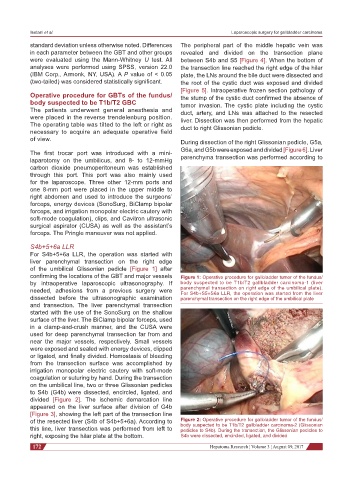
S4b+5+6a LLR
For S4b+5+6a LLR, the operation was started with
liver parenchymal transection on the right edge
of the umbilical Glissonian pedicle [Figure 1] after
confirming the locations of the GBT and major vessels Figure 1: Operative procedure for gallbladder tumor of the fundus/
by intraoperative laparoscopic ultrasonography. If body suspected to be T1b/T2 gallbladder carcinoma-1 (liver
needed, adhesions from a previous surgery were parenchymal transection on right edge of the umbilical plate).
For S4b+S5+S6a LLR, the operation was started from the liver
dissected before the ultrasonographic examination parenchymal transection on the right edge of the umbilical plate
and transection. The liver parenchymal transection
started with the use of the SonoSurg on the shallow
surface of the liver. The BiClamp bipolar forceps, used
in a clamp-and-crush manner, and the CUSA were
used for deep parenchymal transection far from and
near the major vessels, respectively. Small vessels
were exposed and sealed with energy devices, clipped
or ligated, and finally divided. Hemostasis of bleeding
from the transection surface was accomplished by
irrigation monopolar electric cautery with soft-mode
coagulation or suturing by hand. During the transection
on the umbilical line, two or three Glissonian pedicles
to S4b (G4b) were dissected, encircled, ligated, and
divided [Figure 2]. The ischemic demarcation line
appeared on the liver surface after division of G4b
[Figure 3], showing the left part of the transection line
of the resected liver (S4b of S4b+5+6a). According to Figure 2: Operative procedure for gallbladder tumor of the fundus/
body suspected to be T1b/T2 gallbladder carcinoma-2 (Glissonian
this line, liver transection was performed from left to pedicles to S4b). During the transection, the Glissonian pedicles to
right, exposing the hilar plate at the bottom. S4b were dissected, encircled, ligated, and divided
172 Hepatoma Research ¦ Volume 3 ¦ August 09, 2017