
Page 138 - Read Online
P. 138
Zhu et al. Function of Mcl-1 and its regulative relations with HCC
advances in the relationship between HCC treatment that alternative splicing occurred in the transcription of
and anti-apoptotic molecule Mcl-1 and suggests that Mcl-1 and eventually generated 2 different transcript
Mcl-1 is a potential target in abolishing the HCC cells’ variants. The one including 3 exons encodes Mcl-1L
malignant proliferation. isoform while the other lack of exon 2 encodes Mcl-
1S isoform. Sequence analysis revealed that Mcl-1L
Bcl-2 is a well established family of proteins and contains 350 residues which is larger than Bcl-2 (237
has a significant impact on mitochondrial integrity residues) and Bcl like protein X (Bcl-xl) (233 residues)
by influencing the permeability of the mitochondrial and has 3 homo domains BH1, BH2, BH3 and C-terminal
membrane. Bcl-2 is localized to the outer membrane transmembrane (TM) domains but lack the N-terminal
of mitochondria, where it plays a part by regulating the BH4 domain compared to Bcl-2 and Bcl-xl. The TM
progression of apoptosis. According to the structures domain could anchor Mcl-1L to the outer mitochondrial
and the functional contribution, Bcl-2 family members membrane (OMM). [14] By contrast, Mcl-1S comprises
can be divided into two subfamilies: pro-apoptotic 271 residues and retains only BH3 domains just like
members and anti-apoptotic members. And it is other BH3-only members of Bcl-2 family and is primarily
[5]
the balance in activity between the two opposing localized to the cytosol. Surprisingly, Mcl-1L inhibits
groups which determines a cell’s progression towards apoptosis while Mcl-1S exhibits an opposite role and
apoptosis. promotes apoptosis. [13,15] Different from other proteins
of Bcl-2 family, the N-terminal region of Mcl-1 (Mcl-1L
Mcl-1 as an antiapoptotic Bcl-2 family protein, is will be simply called as Mcl-1 hereafter), affecting Mcl-
playing a pivotal role in the intrinsic apoptosis pathway 1’s function and localization, is larger than that of other
and mitotic regulators. As reported, Mcl-1 expresses Bcl-2 family members which contains PEST sequences
[6]
extensively in the normal tissue of human and its rich in proline (P), gluatamic acid (E), serine (S), and
overexpression is observed in many types of human threonine (T). As characteristic sequences of Mcl-1, the
tumors. In addition, Mcl-1 expression involves in PEST regions are rich in putative regulatory motifs that
disease grade and survival in human malignancies have been shown to target proteins for degradation,
e.g. in patients with multiple myeloma or B-cell non- which are thought to be as the main reasons of the
Hodgkin’s lymphoma. [7,8] It is also one of the pervasive short half-life of Mcl-1 protein. [14,16] There are also
recognized anti-apoptosis factor in HCC and mainly multiple phosphorylation sites in Mcl-1 PEST region,
participate in maintenance of mitochondrial membrane and it is likely that multiple proteins resulting in different
stability and suppresses cytochrome c release from fates of Mcl-1 mediate the phosphorylation of these
mitochondria to promote cell survival and inhibit cell sites. Moreover, with a surface-exposed hydrophobic
apoptosis. In addition, Mcl-1, serves as one of the groove formed by BH1, BH2, and BH3, Mcl-1 can
[9]
important antiapoptotic factors in HCC, is involved in integrate with other pro-apoptotic protein containing
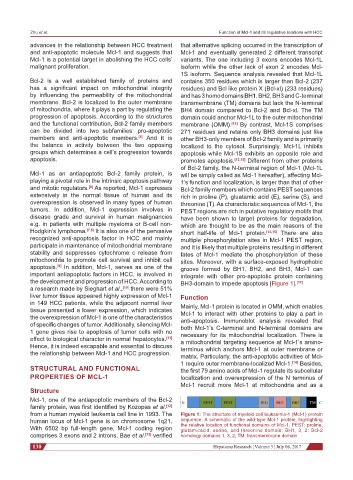
the development and progression of HCC. According to BH3-domain to impede apoptosis [Figure 1]. [17]
a research made by Sieghart et al., [10] there were 51%
liver tumor tissue appeared highly expression of Mcl-1 Function
in 149 HCC patients, while the adjacent normal liver Mainly, Mcl-1 protein is located in OMM, which enables
tissue presented a lower expression, which indicates Mcl-1 to interact with other proteins to play a part in
the overexpression of Mcl-1 is one of the characteristics anti-apoptosis. Immunoblot analysis revealed that
of specific changes of tumor. Additionally, silencing Mcl- both Mcl-1’s C-terminal and N-terminal domains are
1 gene gives rise to apoptosis of tumor cells with no necessary for its mitochondrial localization. There is
effect to biological character in normal hepatocytes. [11] a mitochondrial targeting sequence at Mcl-1’s amino-
Hence, it is indeed escapable and essential to discuss terminus which anchors Mcl-1 at outer membrane or
the relationship between Mcl-1 and HCC progression. matrix. Particularly, the anti-apoptotic activities of Mcl-
1 require outer membrane-localized Mcl-1. [18] Besides,
STRUCTURAL AND FUNCTIONAL the first 79 amino acids of Mcl-1 regulate its subcellular
PROPERTIES OF MCL-1 localization and overexpression of the N terminus of
Mcl-1 recruit more Mcl-1 at mitochondria and as a
Structure
Mcl-1, one of the antiapoptotic members of the Bcl-2
family protein, was first identified by Kozopas et al. [12]
from a human myeloid leukemia cell line in 1993. The Figure 1: The structure of myeloid cell leukaemia-1 (Mcl-1) protein
human locus of Mcl-1 gene is on chromosome 1q21. sequence. A schematic of the wild-type Mcl-1 protein, highlighting
With 6502 bp full-length gene, Mcl-1 coding region the relative location of functional domains of Mcl-1. PEST: proline,
glutamicacid, serine, and threonine domain; BH1, 3, 2: Bcl-2
comprises 3 exons and 2 introns. Bae et al. [13] verified homology domains 1, 3, 2; TM: transmembrane domain
130 Hepatoma Research ¦ Volume 3 ¦ July 06, 2017