
Page 134 - Read Online
P. 134
El-Gazzar et al. Gadobenate dimeglumine dynamic MRI for early HCC detection
Table 3: Combinations of different phases of MRI group of atypical HCC selected is a challenging group
Phase HCC patients Non-HCC patients for diagnosis. Guidelines for diagnosis of atypical
Arterial phase hyperintensity 7 4 cases of HCC are not yet well established and need
HBP hypointense further studies using different imaging modalities for
Hypovascular, HBP 1 7 early diagnosis thereby optimizing treatment outcome.
hypointense
Arterial phase hyperintensity 2 1
HBP hypointense, rim In our study, 8 out of 30 cirrhotic patients with atypical
enhancement hepatic focal lesions on triphasic CT, were diagnosed
HCC: hepatocellular carcinoma; HBP: hepatobiliary phase; MRI: as HCC based on positive immunostaining of at least
magnetic resonance imaging
2 HCC biomarkers (GLP3, HSP70, or GS) according
patients and 1 non-HCC patient [Table 2]. to the international consensus group for hepatocellular
neoplasia. Seven out of these 8 lesions were
On combining different phases, it was found that diagnosed by gadobenate dimeglumine-enhanced MRI
5 HCC patients showed T2 hyper-intensity and (multihance) as HCC, showing hyperintensity in the
hypervscular HBP hypointense lesions, those findings arterial phase and hypointensity in the HBP (according
were not detected in non-HCC patients. Seven non- to the latest guidelines for diagnosis of HCC including
HCC patients showed hypovascular HBP hypointense those of the Japan Society of Hepatology (JSH), [12]
lesions versus 1 HCC patient [Table 3]. Two HCC the Korean Liver Cancer Study Group (KLCSG),
patients showed hypervascular HBP hypointense and the National Cancer Center (NCC), [13] and the Liver
rim enhancement at delayed phase versus 1 non-HCC Imaging Reporting and Data System (LI-RADS). [14]
patient [Table 3]. The sensitivity on combining the
arterial phase hyperintensity and HBP hypointensity According to the updated 2014 JSH guidelines, non-
was 87.5% and specificity was 82.8%. invasive diagnosis of HCC can be made using a
hepatobiliary contrast if a lesion shows: (1) arterial
DISCUSSION hypervascularity and venous washout or (2) arterial
hypervascularity without venous washout, but with
Gadobenate dimeglumine is not a novel agent but the hypointensity on the HBP. [15]
A B
C D
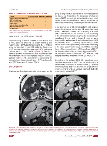
Figure 3: Trans-axial magnetic resonance images of the liver demonstrates right lobe focal lesion (arrow). It appears hyper-intense on T2
image (A). The arterial phase (B) shows faint enhancement in the lesion with washout on the arterio-portal phase (C) and the appearance of
capsule. The lesion is hypo-intense to the liver parenchyma on the hepato-biliary phase (D). This lesion proved to hepatocellular carcinoma
on histopathology
126 Hepatoma Research ¦ Volume 3 ¦ June 26, 2017