
Page 133 - Read Online
P. 133
El-Gazzar et al. Gadobenate dimeglumine dynamic MRI for early HCC detection
Table 1: Patient and tumor characteristics Table 2: MRI with multihance findings in all patients
Variable Number Finding description HCC patients Non-HCC patients
Mean diameter of nodules (cm) Hypointenisity on T1 4 5
HCC 2.1 Hyperintensity on T2 5 6
Non-HCC 1.2 Arterial hyperintensity 7 7
Laboratory tests Delayed hypointensity 0 2
ALT (U/L) 35 ± 28 Hepatobiliaryhypointensity 7 5
AST (U/L) 45 ± 30 Rim enhancement 2 1
Total bilirubin (mg/dL) 1.0 ± 0.5
Albumin (g/dL) 4.0 ± 0.7 HCC: hepatocellular carcinoma; MRI: magnetic resonance imaging
INR 1.2 ± 0.2
9
Platelets( x10 /L) 135 ± 80 showed that only 8 patients (26.6%) had at least 2
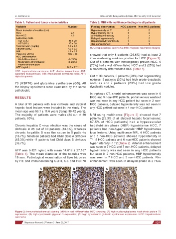
HCC differentiation immunostaining markers positive for HCC [Figure 2].
Well differentiated 6 (75%)
Moderately differentiated 2 (25%) Out of 8 patients with histologically proven HCC, 6
Poorly differentiation (75%) had a well differentiated HCC and 2 (25%) had
AFP (ng/mL) 14.6 ± 27.1 a moderately differentiated HCC [Table 1].
HCC: hepatocellular carcinoma; ALT: alanine transaminase; AST:
aspartate transaminase; INR: international normalized ratio; AFP:
alpha fetoprotein Out of 30 patients, 6 patients (20%) had regenerating
nodules, 9 patients (30%) had high grade dysplastic
70 (HSP70) and glutamine synthetase (GS). All nodules and 7 patients (23%) had low grade
the biopsy specimens were examined by the same dysplastic nodules.
pathologist.
In triphasic CT, arterial enhancement was seen in 6
RESULTS HCC and 5 non-HCC patients, portal venous washout
was not seen in any HCC patient but seen in 2 non-
A total of 30 patients with liver cirrhosis and atypical HCC patients, delayed hypointensity was not seen in
hepatic focal lesions were included in the study. The any HCC patient but seen in 1 non-HCC patient.
mean age was 56.1 ± 11.6 years (range 39-72 years).
The majority of patients were males (24 out of 30 MRI using multihance [Figure 3] showed that 7
patients, 80%). patients (23.3% of all atypical hepatic focal lesions;
87.5% of HCC patients) had a hypevascular
Chronic hepatitis C virus infection was the cause of hepatobiliary phase (HBP) hypointense HCC, 7
cirrhosis in 25 out of 30 patients (83.3%), whereas patients had non-hyper vascular HBP hypointense
chronic hepatitis B was the cause in 5 patients focal lesions. Using multihance MRI, 4 HCC patients
(16.7%). Nineteen patients had Child class A cirrhosis and 5 non-HCC patients showed hypointensity in
(63.3%) while 11 patients had Child class B cirrhosis T1, 5 HCC patients and 6 non-HCC patients showed
(36.7%). hyper intensity in T2 [Table 2]. Arterial enhancement
was seen in 7 HCC and 7 non-HCC patients, delayed
AFP was 8-121 ng/mL with mean 14.619 ± 27.187 hypointensity was not seen in any HCC patients
[Table 1]. The mean diameter of the nodules was but seen in 2 non-HCC patients, HBP hypointensity
19 mm. Pathological examination of liver biopsies was seen in 7 HCC and 5 non-HCC patients. Rim
by HE and immunostaining GLP3, GS and HSP70 enhancement was seen in delayed phase in 2 HCC
A B C
Figure 2: Immunohistochemical staining (x400) in well differentiated HCC showing: (A) high nucleocytoplasmic heat shock protein 70
expression; (B) high cytoplasmic glypican 3 expression; (C) high cytoplasmic glutamin synthetase expression. HCC: hepatocellular
carcinoma
Hepatoma Research ¦ Volume 3 ¦ June 26, 2017 125