
Page 139 - Read Online
P. 139
Zhu et al. Function of Mcl-1 and its regulative relations with HCC
related to the ratio of Mcl-1/Mcl-1S. [23] It is noteworthy
BH3 only Mcl-1 that Mcl-1 plays the leading role in the regulation of
proteins apoptosis induced by Mcl-1/Mcl-1S and is expressed
Direct Indirect
activation activation at higher levels than Mcl-1S. [13,24,25] In cancer, Mcl-1S
is expressed at much lower levels than Mcl-1 that it
Bak/Bax was even hardly undetectable. [26] Some cancer cells
Cytochrome c release such as human lung cancer cell lines A549, Chinese
hamster ovary cells and multiple myeloma MOLP-
Apoptosome [13,15,27]
formation 8 cells show high level of Mcl-1S. Hence, this
review mainly discusses Mcl-1.
Caspase
activation Paradoxically, it is possible that Mcl-1 also plays an
important role in delaying cell cycle progression for the
existence of Mcl-1 in nucleus have been reported as
well. [28] The first 79 amino acids of Mcl-1 promotes its
Apoptosis association with mitochondria, the N terminus of Mcl-1
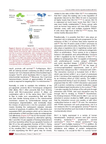
Figure 2: Myeloid cell leukaemia-1 (Mcl-1) regulates initiation of also plays a regulatory role in regulating nuclear (anti-
apoptosis through interaction with other Bcl-1 family members. proliferative) functions of Mcl-1 and has an antagonistic
Under normal circumstances, Mcl-1 prevents the activation of Bax effect on proliferation. There seems to be a balance
and Bak to protect mitochondrial outer membrane integrity and
cell survival. Under conditions of stress, the BH3 only proteins between anti-apoptotic and anti-proliferative functions
are activated and induce apoptosis either by releasing Bak/Bax of Mcl-1 regulated by the N terminus of Mcl-1. [19] In
from Mcl-1 or by BH3 only proteins binding to Mcl-1 directly. Bak/
Bax form pores on mitochondrial outer membrane and cytochrome addition to antiapoptosis, Mcl-1 is capable of interacting
C is relieved into the cytoplasm. In the cytoplasm, cytochrome C with proliferating-cell nuclear antigen (PCNA) [29]
activates a family of cysteine proteases named caspases which and cyclin depndent kinase 1 (CDK1), [4] which may
subsequently induce cell apoptosis
inhibit cell cycle progression. [29,30] On the basis of
result, promote cell survival. [19] Furthermore, Mcl-1 co-immunoprecipitation experiments, Jamil et al. [30]
with an internal domain containing an EELD motif (at showed that endogenous Mcl-1 interacted with CDK1.
residue 124-127) interacts with the mitochondrial import The interaction involved a truncated form of Mcl-1,
receptor Tom70, which facilitates Mcl-1’s import onto which was termed snMcl-1 as a result of proteolysis
mitochondrial membrane. [20] Moreover, the C-terminal at the C-terminus that regulated cell-cycle progression
transmembrane domain of approximately 20 amino by an inhibitory effect on CDK1 activity. The snMcl-1
acids is required for targeting Mcl-1 to mitochondria. [21] was presented during S and G2 phases. The authors
proposed that the Mcl-1-CDK1 interaction associated
Normally, in order to maintain the inhibition of the with a protein containing a nuclear localization signal
pro-apoptotic proteins Bcl-2 homologous antagonist that mediated rapid translocation to the nucleus. [30] Mcl-
killer (Bak), Mcl-1 often prevents Bak from forming 1 can also regulate the S-phase of the cell cycle through
dimer with Bcl-2-associated protein X (Bax) via interaction with PCNA, and such interaction may be
combination with it. At the same time, Mcl-1 binds through Mcl-1’s binding to PCNA. [29] Nonetheless,
to Bax to make sure sequestering Bak and Bax, the binding between PCNA and the Mcl-1 can not be
and then blocks forming pores in the mitochondrial detected in solution studied by NMR, which suggests
membrane caused by conformational change and that the interaction occurs very weakly, or with other
homologous oligomerization, and eventually stops unidentified factors in cells. [31] Of note, the interaction
the release of cytochrome c into the cytoplasm, which with PCNA represses cell cycle progression, but it is
means blocking the subsequent caspase cascade not related to Mcl-1’s anti-apoptotic activity. [29] And it’s
reaction of apoptosis. In addition, Mcl-1 also binds and not clear that whether such two kinds of interaction are
sequesters BH3 only proteins which act to induce the mechanistically linked.
polymerisation of Bak and Bax to play its antiapoptosis
role effectively [Figure 2]. [22] As for the function of Mcl- Mcl-1 is highly expressed in a variety of human
1’s different isoforms induced by alternative splicing, hematopoietic, lymphoid cancers and solid tumors
Mcl-1 and Mcl-1S are capable of forming heterodimers including leukemia, [32,33] lymphoma, [8] cervical
and thus neutralize either the pro-apoptotic function of carcinoma, [34] HCC, [10,35] breast carcinoma, [36] lung
Mcl-1S or the anti-apoptotic function of Mcl-1. Owing cancer [37] and multiple myeloma. [7,38,39] In addition, its
to alternative splicing mechanisms and interactions expression is often implicated in the chemotherapeutic
of the resulting Mcl-1 and Mcl-1S proteins, the fate resistance and relapse of certain malignancies.
of cells expressing the Mcl-1 gene may be closely For instance, it is crucial for Mcl-1 to survive human
Hepatoma Research ¦ Volume 3 ¦ July 06, 2017 131