
Page 140 - Read Online
P. 140
Zhu et al. Function of Mcl-1 and its regulative relations with HCC
myeloma cells in vitro and it has been showed that Mcl- activators of transcription (STAT), cAMP-response
1 is overexpressed in vivo in multiple myeloma, which element binding protein (CREB), purine-rich nucleic acid
seems to be related to relapse and shorter survival. binding protein 1 (PU.1), and hypoxia-inducible factor-1
[7]
Expression of Mcl-1 was also bound up with high tumor (HIF-1), etc. The STATs, a family of transcription factors,
grade and reduced survival of patient in human breast has been shown to bind to Mcl-1 promoter. Al Zaid
cancer samples. [40] Immunohistochemistry and western Siddiquee and Turkson [46] reported that constitutively
blotting analysis showed that Mcl-1 was overexpressed activated STAT3 participate in oncogenesis of the liver
in cervical cancer tissue in comparison with normal through up-regulating STAT3-targeted genes encoding
tissue and the author confirmed Mcl-1 expression apoptosis inhibitors including Mcl-1 and subsequently
was positively correlated with poor prognosis. [34] As inhibiting pro-apoptotic molecules such as Bax, Bad,
for acute myeloid leukemia, Mcl-1 served as a critical and Bid. Additionally, sorafenib was affirmed for its
molecule to develop and maintain malignant tumor. [41,42] efficacy against Janus Kinase (JAK)-STAT signaling in
Moreover, Campbell et al. [43] reported that elevated HCC cells and downregulation of pSTAT3 and its target
Mcl-1 promotes Myc-induced lymphomagenesis and genes including Mcl-1 by immunblotting. [47] Irophic
enhances drug resistance. And also, in human HCC, factor IL-3 also involves in transcriptional upregulation
it has been concluded that Mcl-1 expression was of Mcl-1. Through activation of the PU.1 transcription
prominently enhanced in diseased tissue as well as in factor, IL-3 activates Mcl-1 transcription by the P38
various HCC cell lines. [10,35] On the contrary, in mice mitogen-activated protein kinase (MAPK)-dependent
lacking the anti-apoptotic protein Mcl-1 specifically in pathway. [48] On the other hand, Mcl-1 transcription
hepatocytes not only increased hepatocyte apoptosis, can also be activated by IL-3 increasing of the DNA
but also resulted in hepatocarcinogenesis, which is binding activity of the CRE-2 binding complex through
related to compensatory hyper-proliferation induced phosphatidylinositol-3-kinase (PI3K)/Akt signaling
by Mcl-1 deficiency. [44] Besides, another mouse pathway. [49] HIF-1 is a putative key transcription factor
model indicates that Mcl-1 is stabilized by interleukin which can regulate cells under hypoxia undergoing
(IL)-6 and obesity and thus apoptosis of damaged different transcriptional adaptations. [50] Through
hepatocytes was inhibited, which eventually promoted analysis of the Mcl-1 promoter sequence in hepatoma
HCC progression. [45] HepG2 cells incubated under hypoxia, Jean-Pascal
Piret et al. [51] demonstrated that there was a hypoxia-
REGULATIVE RELATIONS WITH HCC responsive element in Mcl-1 promoter fragment
that was able to bind HIF-1 in vitro. Detailed results
Combining unrestrained cell proliferation and damaged revealed that HIF-1 showed a potential anti-apoptotic
apoptosis was found as a main feature of tumor. And role and could protect cells against apoptosis as
as mentioned before, the anti-apoptotic member Mcl- a result of hypoxia by up-regulation of the Mcl-1
1 was overexpressed in HCC endowing tumor cells protein. [51] Luciferase reporter assay revealed that
with ability to escape from programmed cell death. overexpression of periostin enhanced HIF-1α–
Consequently, it is of great necessary to make clear dependent transcriptional activity and induced multiple
the regulation and execution of apoptosis in HCC so HIF-1α target genes including Mcl-1, and Bcl-xL in
that people can find a new way to confront malignant HCC cells. [52] Moreover, the ternary complex factor-
tumor. The regulative relation between Mcl-1 and HCC serum response factor complex are also involved in
is listed in Table 1. regulating Mcl-1 expression and protecting cells from
apoptotic cell death. [53] After activating cells with a
Transcriptional regulation variety of cytokines, Mcl-1 expression can be regulated
Mcl-1 can be regulated at transcriptional level by a transcriptionally in several signaling pathways. A recent
variety of cytokines including signal transducers and report describes that after treatment of HCC SK-Hep-1
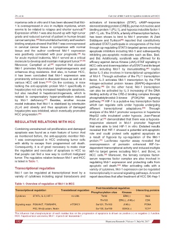
Table 1: Overview of regulation of Mcl-1 in HCC
Post-translational regulation
Transcriptional regulation Translational regulation Interacting proteins
Phosphorylation sites Kinases
Cytokines STATs, IL-3, HIF-1 mir-29b Thr92 ERK(-) Mule
Thr163 ERK(-), JNK(+) CDK
Signaling ways PI3K/AKT, P38/MAPK, mTORC1 Ser121, Thr163 JNK(-) PCNA
P53, Wnt/β-actein, Notch Ser155, Ser159 GSK3(+) TCTP
The influence that phosphorylation of each residue has on the progression of apoptosis is shown as positive (+) or negative (-) function.
HCC: hepatocellular carcinoma; Mcl-1: myeloid cell leukaemia-1
132 Hepatoma Research ¦ Volume 3 ¦ July 06, 2017