
Page 142 - Read Online
P. 142
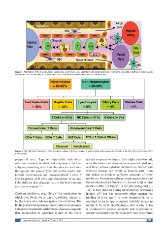
Figure 1: Architecture of the liver: sinusoids, hepatocytes and immune cells. LSEC form a fenestrated monolayer within the sinusoidal endothelium. HSC: hepatic
            stellate cells; NK: natural killer; KC: Kupffer cells; LSEC: liver sinusoidal endothelial cells; DC: dendritic cells


            Figure 2: The estimated frequency of each population relative to the total number of parenchymal and nonparenchymal cells in the liver. NK: natural killer; TCR:
            T-cell receptor
            periportal area.  Together  sinusoidal endothelial   natural response to illness. One might therefore ask
            cells and resident dendritic cells represent the liver   what the balance is between the amount of cytokines
            antigen presenting cells. Lymphocytes are scattered   and these natural cytokine inhibitors in disease and
            throughout the parenchyma and portal tracts, and   whether  disease  can result,  at  least  in  part, from
            include conventional and unconventional T cells. A   the  failure  to  produce sufficient  amounts  of  these
            low frequency of B cells and abundance of natural   inhibitors. For instance, the principle agonist forms of
            killer (NK) are also characteristic of the liver immune   the interleukin (IL)-1 family are IL-1α and IL-1β. A third
            microenvironment. [13]                             member of the IL-1 family, IL-1 receptor antagonist (IL-
                                                               1ra), is also induced during inflammatory responses
            Cytokine inhibitors, regardless of the mechanism by   [Figure 3]  but has preventive  effect against  the
                                                                        [14]
            which they block the action of cytokines, appear to   binding of IL-1α and  β to their receptors  IL-1ra is
            be the host’s own defense against the cytokines. The   required to be in approximately 100-fold excess to
            finding of elevated plasma concentrations of cytokines   inhibit IL-1α or IL-1β effectively; that is why IL-1ra
            antagonist in patients with various diseases suggests   is  produced in greater amounts and is present  at
            that antagonism  to cytokines is part of the host’s   greater concentration matched with rare occurrence


                 Hepatoma Research | Volume 2 | June 1, 2016                                              133