
Page 113 - Read Online
P. 113
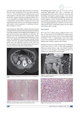
scan with contrast and triple phase abdomen CT scan were   The pathology report showed a 6.5 cm × 6.1 cm × 6.0 cm
           done for better visualization of the mass which showed a   moderately differentiated HCC with a trabecular and
           7.2 cm × 5.8 cm heterogeneous enhancing mass in the   pseudoglandular growth pattern with foci of necrosis and
           sixth segment of the liver, with a pseudocapsule bulges   hemorrhage and negative surgical margins, it also showed
           on the liver capsule, which has an adjacent small 6 cm ×   vascular invasion of the portal triad and diffuse macronodular
           3 cm accumulation of complex fluid that likely represents   cirrhosis [Figures 3 and 4]. At this point, the decision was
           a ruptured HCC and less likely a benign liver tumor with   made to follow-up the patient closely with AFP, liver function
           regional hematoma, the liver was abnormal in appearance   test, and imaging studies every 3 months for the first 2 years.
           with nodular contour suggestive of underlying cirrhosis/  Follow-up AFP about 1 month after surgery was 54.4 ng/mL
           fibrosis [Figures 1 and 2].                        and 4.8 ng/mL after 3 months.

           Gastroenterology, hepatobiliary surgery, and interventional   DISCUSSION
           radiology (IR) were consulted, after that and while the patient
           was being evaluated, her hemoglobin level dropped to 12.2   HCC is the most common primary malignant tumor of the
           g/dL and she became more tachycardic. At that time, the   liver; it is also known to be the fifth most common cancer
           impression was that the patient has a ruptured HCC, and the   and the third most common cause of cancer-related death
           decision was to do an IR-guided bland embolization of the   worldwide.   HCC  is  a  hypervascular  tumor  that  mostly
                                                                       [1,2]
           tumor. The embolization was done using polyvinyl alcohol   occurs in the settings of liver cirrhosis, and it is one of the
           particles. After that, the patient remained hemodynamically   leading causes of death in patients with cirrhosis.
           stable. Later, she underwent resection of the tumor and
           the sixth segment of the liver, during surgery there was no   Spontaneous rupture is a major life-threatening complication
           evidence of spread of the tumor outside the liver.  of HCC that occurs in 3-15% of cases with geographical
                                                              differences  among  Western,  Asian,  and  African  countries,
                                                              where HCC is more frequent.  The incidence of HCC is on
                                                                                     [3]
                                                              decline due to early detection and screening.


                                                              Figure 2: Computed tomography scan confirming evidence of hepatocellular
           Figure 1: Computed tomography scan revealing evidence of hepatocellular   cancer with regional hematoma
           cancer


           Figure 3: Histopathology of liver tissue revealing a trabecular and
           pseudoglandular growth pattern with foci of necrosis and hemorrhage  Figure 4: Histopathology of resected hepatic tumor

           104                                                            Hepatoma Research | Volume 2 | April 1, 2016