
Page 80 - Read Online
P. 80
Page 6 of 8 Ban et al. Hepatoma Res 2021;7:13 I http://dx.doi.org/10.20517/2394-5079.2020.104
A
B
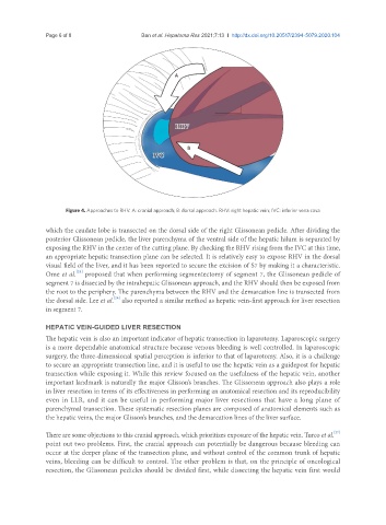
Figure 4. Approaches to RHV. A: cranial approach; B: dorsal approach. RHV: right hepatic vein; IVC: inferior vena cava
which the caudate lobe is transected on the dorsal side of the right Glissonean pedicle. After dividing the
posterior Glissonean pedicle, the liver parenchyma of the ventral side of the hepatic hilum is separated by
exposing the RHV in the center of the cutting plane. By checking the RHV rising from the IVC at this time,
an appropriate hepatic transection plane can be selected. It is relatively easy to expose RHV in the dorsal
visual field of the liver, and it has been reported to secure the excision of S7 by making it a characteristic.
[25]
Ome et al. proposed that when performing segmentectomy of segment 7, the Glissonean pedicle of
segment 7 is dissected by the intrahepatic Glissonean approach, and the RHV should then be exposed from
the root to the periphery. The parenchyma between the RHV and the demarcation line is transected from
[26]
the dorsal side. Lee et al. also reported a similar method as hepatic vein-first approach for liver resection
in segment 7.
HEPATIC VEIN-GUIDED LIVER RESECTION
The hepatic vein is also an important indicator of hepatic transection in laparotomy. Laparoscopic surgery
is a more dependable anatomical structure because venous bleeding is well controlled. In laparoscopic
surgery, the three-dimensional spatial perception is inferior to that of laparotomy. Also, it is a challenge
to secure an appropriate transection line, and it is useful to use the hepatic vein as a guidepost for hepatic
transection while exposing it. While this review focused on the usefulness of the hepatic vein, another
important landmark is naturally the major Glisson’s branches. The Glissonean approach also plays a role
in liver resection in terms of its effectiveness in performing an anatomical resection and its reproducibility
even in LLR, and it can be useful in performing major liver resections that have a long plane of
parenchymal transection. These systematic resection planes are composed of anatomical elements such as
the hepatic veins, the major Glisson’s branches, and the demarcation lines of the liver surface.
[27]
There are some objections to this cranial approach, which prioritizes exposure of the hepatic vein. Turco et al.
point out two problems. First, the cranial approach can potentially be dangerous because bleeding can
occur at the deeper plane of the transection plane, and without control of the common trunk of hepatic
veins, bleeding can be difficult to control. The other problem is that, on the principle of oncological
resection, the Glissonean pedicles should be divided first, while dissecting the hepatic vein first would