
Page 77 - Read Online
P. 77
Ban et al. Hepatoma Res 2021;7:13 I http://dx.doi.org/10.20517/2394-5079.2020.104 Page 3 of 8
A B C
D E
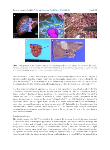
Figure 1. Laparoscopic partial liver resection of segment 5. A: preoperative simulation; B: the resection plan is to include the MHV on
the dorsal side of the tumor on the resection side. White arrows indicate MHVs; C: the targeted MHV is encircled on the left side of the
resection plane; D: the planned MHV is included on the resection side; E: the divided surface of the specimen after resection. The white
arrow indicates the MHV, which is useful as an indicator of the resection margin
the confluence of the veins does not split. In addition, the viewing angle when laparoscopic surgery is
performed differs from that of open surgery and has the angular characteristics of approaching the vein
[11]
from the dorsal side . If the working site of the hepatectomy is on the ventral side, the tiny amount of
[10]
bleeding produced will flow to the dorsal side, keeping the area where the operation is performed dry .
Another major advantage of laparoscopic surgery is that laparoscopic magnification allows for the
observation of detailed anatomy. Reports on liver resection focusing on Laennec’s capsule have recently
been published . Particularly during dissection of the hepatic vein near the inflow of the vein into the
[12]
inferior vena cava (IVC), it is useful to be aware of Laennec’s capsule and to select the layers of the vein
[13]
during dissection, as any injury can be critical . Kiguchi et al. reported that the layer between the
[14]
hepatic and cardiac Laennec’s capsule around the root of the hepatic veins could be divided and the hepatic
veins safely exposed. He advocated an “inter-Laennec approach” that enables liver dissection preserving
[15]
only the cardiac Laennec’s capsule around the vein wall . This approach might have the advantage of
securing the resection margin when the tumor is close to the hepatic vein. The role of the major hepatic
veins is described below.
Middle hepatic vein
The middle hepatic vein (MHV) is located in the center of the liver, and thus, it is the most important
landmark and has a wide range of applications. It runs along the area boundary between the right and
left lobes of the liver and is located in a discrete section of the Cantlie line. As a matter of course, it runs
along the transection plane of the right or left lobectomy. The MHV serves as a landmark on the right
side of the transection plane for left internal sectionectomy and on the left side of the dissected section
for right anterior sectionectomy. In addition, during the segmentectomy of S8, when the MHV is exposed
from the central side, the Glisson branch of S8 can be identified so as to cross MHV as the method of the