
Page 62 - Read Online
P. 62
Page 6 of 14 Schwertheim et al. Hepatoma Res 2020;6:41 I http://dx.doi.org/10.20517/2394-5079.2020.23
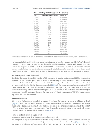
Table 2. NGS study: CTNNB1 mutations in the HCC cohort 1
Gene AA mutation Cosmic_v70 n cases (%)
CTNNB1 2 D32G 1/72 (1.4%)
CTNNB1 2 D32N 1/72 (1.4%)
CTNNB1 2 D32V 0/72 (0%)
CTNNB1 2 D32Y 3/72 (4.2%)
CTNNB1 S33A 1/72 (1.4%)
CTNNB1 2 S33C 3/72 (4.2%)
CTNNB1 2 S33F 2/72 (2.8%)
CTNNB1 2 G34E 1/72 (1.4%)
CTNNB1 G34R 1/72 (1.4%)
CTNNB1 2 G34V 1/72 (1.4%)
CTNNB1 I35S 1/72 (1.4%)
CTNNB1 H36P 1/72 (1.4%)
CTNNB1 S37A 1/72 (1.4%)
CTNNB1 2 S37Y 1/72 (1.4%)
CTNNB1 2 T41A 1/72 (1.4%)
CTNNB1 2 T41I 2/72 (2.8%)
CTNNB1 2 S45P 2/72 (2.8%)
CTNNB1 2 S45Y 1/72 (1.4%)
CTNNB1 K335T 1/72 (1.4%)
CTNNB1 N387K 2/72 (2.8%)
2
1 All mutations found in COSMIC with the prevalence of minimum 5% are listed; mutations listed additionally in the ClinVar database
intranuclear inclusion with positive immunoreactivity was analyzed for β-catenin and KDM2A. We detected
in 19 of 72 (26.4%) HCCs at least one membrane-bounded intranuclear inclusion with positive β-catenin
immunostaining; for KDM2A 19 of 71 (26.8%) valid HCC cases showed at least one membrane-bounded
intranuclear inclusion with positive immunostaining. β-catenin immunohistochemistry was available for all
cases (72/72) while for KDM2A, suitable material for immunohistochemistry was available in 71 of 72 cases.
NGS study of CTNNB1 mutations
To clarify the reason for the high number of NI containing β-catenin, we investigated HCCs with possible
mutations of the β-catenin gene CTNNB1 by NGS. We found up to twenty different CTNNB1 mutations in
our cases. We have listed all mutations found in COSMIC with a prevalence of at least 5%. The mutations that
are also recorded in the ClinVar database are marked [Table 2]. Chi-square cross table analysis of the HCC
cases demonstrated that a positive CTNNB1 mutation status was significantly associated with the occurrence
of positive nuclear β-catenin immunostaining (P ≤ 0.001). Additionally, we performed cross table analysis
to examine a possible association between the occurrence of NI and the presence of CTNNB1 mutations; no
significant association was found.
TEM analysis of NI
We performed ultrastructural analysis in order to investigate the content and shape of NI in more detail
[Figure 2]. Our TEM studies showed that NI in HCC in most cases was completely enclosed by the nuclear
membrane. NI contained degenerated cell material, lysosomes and heterolysosomes. In general, the content
of the inclusions had a higher electron density than the cytoplasm, suggesting that NI are not simply passive
invaginations of the cytoplasm into the nucleus [Figure 2].
Immunohistochemical analysis of NI
Association of β-catenin with autophagy-associated proteins in NI
We analysed NI by immunohistochemistry to clarify whether there was an association between the
occurrence of intranuclear inclusions with β-catenin immunopositivity and autophagy [Figure 3]. Recently,
we have examined the autophagy-associated proteins p62, ubiquitin, LC3B, cathepsin B and cathepsin D