
Page 61 - Read Online
P. 61
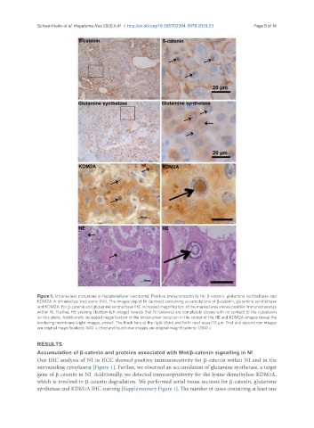
Schwertheim et al. Hepatoma Res 2020;6:41  I  http://dx.doi.org/10.20517/2394-5079.2020.23                                   Page 5 of 14


               Figure 1. Intranuclear inclusions in hepatocellular carcinoma. Positive immunoreactivity for β-catenin, glutamine synthethase and
               KDM2A in intranuclear inclusions (NI). The images depict NI (arrows) containing accumulations of β-catenin, glutamine synthethase
               and KDM2A. For β-catenin and glutamine synthethase IHC increased magnification of the marked area shows positive immunostainings
               within NI. Further, HE-staining (bottom-left image) reveals that NI (arrows) are completely closed with no contact to the cytoplasm
               on this plane. Additionally increased magnification of the intranuclear inclusion in the center of the HE and KDM2A-images reveal the
               bordering membrane (right images; arrow). The black bars at the right (third and forth row) equal 10 µm. First and second row images
               are original magnifications: 400 ×; third and fourth row images are original magnifications: 1,000 ×

               RESULTS
               Accumulation of β-catenin and proteins associated with Wnt/β-catenin signalling in NI
               Our IHC analysis of NI in HCC showed positive immunoreativity for β-catenin within NI and in the
               surrounding cytoplasma [Figure 1]. Further, we observed an accumulation of glutamine synthetase, a target
               gene of β-catenin in NI. Additionally, we detected immunopositivity for the lysine demethylase KDM2A,
               which is involved in β-catenin degradation. We performed serial tissue sections for β-catenin, glutamine
               synthetase and KDM2A IHC staining [Supplementary Figure 1]. The number of cases containing at least one