Page 59 - Read Online
P. 59

Schwertheim et al. Hepatoma Res 2020;6:41  I  http://dx.doi.org/10.20517/2394-5079.2020.23                                   Page 3 of 14


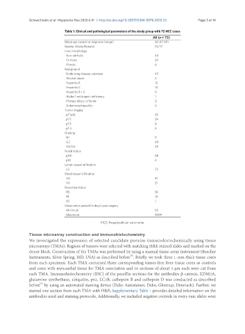
                                Table 1. Clinical and pathological parameters of the study group with 72 HCC cases
                                                                           All (n = 72)
                                Mean age (years) at diagnosis (range)       62 (17-99)
                                Gender (Male/Female)                        55/17
                                Liver morphology
                                  Non-cirrhotic                             44
                                  Cirrhotic                                 22
                                  Fibrotic                                  6
                                Background
                                  Underlying disease unknown                43
                                  Alcohol abuse                             2
                                  Hepatitis B                               12
                                  Hepatitis C                               14
                                  Hepatitis B + C                           0
                                  Alpha-1-antitrypsin deficiency            1
                                  Primary biliary cirrhosis                 0
                                  Autoimmunhepatitis                        0
                                Tumor staging
                                  pT1a/b                                    35
                                  pT2                                       24
                                  pT3                                       8
                                  pT4                                       5
                                Grading
                                  G1                                        9
                                  G2                                        40
                                  G3/G4                                     23
                                Nodal status
                                  pN0                                       68
                                  pN1                                       4
                                Lymph vessel infiltration
                                  L0                                        72
                                Blood vessel infiltration
                                  V0                                        41
                                  V1                                        31
                                Resection status
                                  R0                                        61
                                  R1                                        10
                                  R2                                        1
                                Observation period (in days) post surgery
                                  Minimum                                   55
                                  Maximum                                   3009

                                                  HCC: Hepatocellular carcinoma

               Tissue microarray construction and immunohistochemistry
               We investigated the expression of selected candidate proteins immunohistochemically using tissue
               microarrays (TMAs). Regions of tumors were selected with matching H&E stained slides and marked on the
               donor block. Construction of the TMAs was performed by using a manual tissue-array instrument (Beecher
                                                                [6]
               Instruments, Silver Spring, MD, USA) as described before . Briefly, we took three 1-mm-thick tissue cores
               from each specimen. Each TMA contained three corresponding tumor-free liver tissue cores as controls
               and cores with myocardial tissue for TMA orientation and 10 sections of about 3 µm each were cut from
               each TMA. Immunohistochemistry (IHC) of the paraffin sections for the antibodies β-catenin, KDM2A,
               glutamine synthethase, ubiquitin, p62, LC3B, cathepsin B and cathepsin D was conducted as described
                    [6]
               before  by using an automated staining device (Dako Autostainer, Dako, Glostrup, Denmark). Further, we
               stained one section from each TMA with H&E; Supplementary Table 1 provides detailed information on the
               antibodies used and staining protocols. Additionally, we included negative controls in every run: slides were
   54   55   56   57   58   59   60   61   62   63   64