
Page 65 - Read Online
P. 65
Schwertheim et al. Hepatoma Res 2020;6:41 I http://dx.doi.org/10.20517/2394-5079.2020.23 Page 9 of 14
Table 3. Correlation of β-catenin immunopositivity within NI with autophagy associated proteins in NI and with cytoplasmic β-catenin
Cross Tabs Tumor tissue
Antibody n P value
β-catenin p62 16/72 0.001
LC3B 7/72 0.041
Ubiquitin 15/71 < 0.001
Cathepsin B 17/72 0.001
Cathepsin D 13/72 0.005
Cytoplasmic β-catenin 11/72 0.002
P values were calculated using two-sided Fisher’s exact test. n : Number of IHC positive intranuclear inclusions/valid cases; NI:
intranuclear inclusions
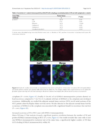
Figure 4. Double-IF studies demonstrate co-localization of β-catenin with p62 in intranuclear inclusions (NI) of hepatocellular
carcinoma sections. Nuclei were stained with DAPI (blue) to detect NI. The merged images show superimposition of the signals for
β-catenin (green staining) with p62 (red staining) in the same inclusion; the merged color yellow (arrow) proves co-localizations
cytoplasm (P = 0.009; Figure 5C); briefly 27 (84.4%) of 32 KDM2A immunopositive patients showed no
local recurrence compared to 7 (46.7%) of 15 patients with lack of KDM2A in the cytoplasm and developed
recurrence. Additionally, we studied the adjacent normal tissue sections (NTS) on all serial sections of the
HCC patient cohort by Kaplan-Meier survival curves. We also detected in the adjacent normal tissue that the
occurrence of KDM2A in the cytoplasm was associated with a significant benefit in recurrence-free survival
(P = 0.027; Figure 5D).
Increased occurrence of NI in HCC cases with KDM2A immunopositivity
Mann-Whitney U Test analysis showed a significant positive correlation between the number of NI and
positive KDM2A immunostaining in HCC (P ≤ 0.001; Figure 6). Our results revealed that cases with at least
one KDM2A immunopositive inclusion had, at the same time, a significantly higher number of NI than
HCCs lacking KDM2A immunoreactivity within NI.