
Page 95 - Read Online
P. 95
Wilson et al. Hepatoma Res 2020;6:57 I http://dx.doi.org/10.20517/2394-5079.2020.48 Page 9 of 11
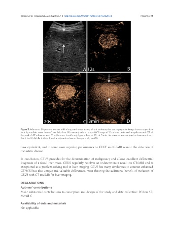
Figure 5. Adenoma. 34-year-old woman with a long continuous history of oral contraceptive use. a greyscale image shows a superficial
focal hypoechoic mass (arrows) in a fatty liver (A); an early arterial phase (AP) image at 12 s shows peripheral irregular vessels (B); at
the peak of AP enhancement, 20 s, the mass is uniformly hyperenhanced (C); at 3 min, the mass shows sustained enhancement such
that it is still slightly brighter than the adjacent enhanced liver parenchyma (D)
have equivalent, and in some cases superior, performance to CECT and CEMR scan in the detection of
metastatic disease.
In conclusion, CEUS provides for the determination of malignancy and allows excellent differential
diagnosis of a focal liver mass. CEUS regularly resolves an indeterminate result on CT/MRI and is
exceptional as a problem-solving tool in liver imaging. CEUS has many similarities to contrast-enhanced
CT/MRI but also unique and valuable differences, most showing the additional benefit of inclusion of
CEUS with CT and MR for liver imaging.
DECLARATIONS
Authors’ contributions
Made substantial contributions to conception and design of the study and date collection: Wilson SR,
Merrill C
Availability of data and materials
Not applicable.