
Page 94 - Read Online
P. 94
Page 8 of 11 Wilson et al. Hepatoma Res 2020;6:57 I http://dx.doi.org/10.20517/2394-5079.2020.48
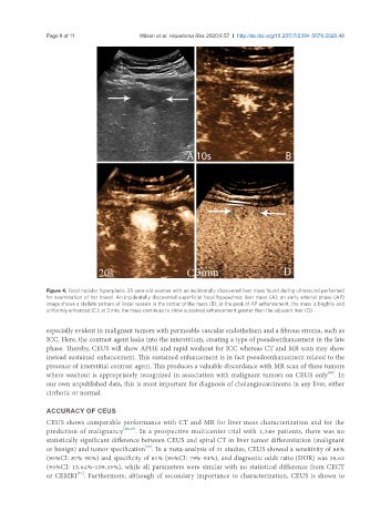
Figure 4. Focal nodular hyperplasia. 25-year-old woman with an incidentally discovered liver mass found during ultrasound performed
for examination of her bowel. An incidentally discovered superficial focal hypoechoic liver mass (A); an early arterial phase (AP)
image shows a stellate pattern of linear vessels in the center of the mass (B); at the peak of AP enhancement, the mass is brightly and
uniformly enhanced (C); at 3 min, the mass continues to show sustained enhancement greater than the adjacent liver (D)
especially evident in malignant tumors with permeable vascular endothelium and a fibrous stroma, such as
ICC. Here, the contrast agent leaks into the interstitium, creating a type of pseudoenhancement in the late
phase. Thereby, CEUS will show APHE and rapid washout for ICC whereas CT and MR scan may show
instead sustained enhancement. This sustained enhancement is in fact pseudoenhancement related to the
presence of interstitial contrast agent. This produces a valuable discordance with MR scan of these tumors
[28]
where washout is appropriately recognized in association with malignant tumors on CEUS only . In
our own unpublished data, this is most important for diagnosis of cholangiocarcinoma in any liver, either
cirrhotic or normal.
ACCURACY OF CEUS
CEUS shows comparable performance with CT and MR for liver mass characterization and for the
prediction of malignancy [26,29] . In a prospective multicenter trial with 1,349 patients, there was no
statistically significant difference between CEUS and spiral CT in liver tumor differentiation (malignant
[30]
or benign) and tumor specification . In a meta-analysis of 21 studies, CEUS showed a sensitivity of 88%
(95%CI: 87%-90%) and specificity of 81% (95%CI: 79%-84%), and diagnostic odds ratio (DOR) was 38.62
(95%CI: 13.64%-109.35%), while all parameters were similar with no statistical difference from CECT
[31]
or CEMRI . Furthermore, although of secondary importance to characterization, CEUS is shown to