
Page 91 - Read Online
P. 91
Wilson et al. Hepatoma Res 2020;6:57 I http://dx.doi.org/10.20517/2394-5079.2020.48 Page 5 of 11
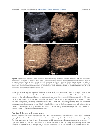
Figure 1. Hepatocellular carcinoma (HCC). 64-year-old male with a history of cirrhosis related to alcohol and fatty liver. Mass found
on regular US surveillance. A greyscale image shows a small hypoechoic liver nodule in a very heterogeneous liver (A); at the peak
of arterial phase (AP) enhancement, 20 s, the mass is brightly enhanced (B); at one minute, there is isoenhancement. The nodule is
now invisible but marked by the arrow (C); there is late and weak washout at 3 min. Weak washout shows less enhancement than the
immediately adjacent liver parenchyma but retains bubble signals within the nodule as here. AP hyperenhancement and late weak
washout comprise the diagnostic features of HCC (D)
technique and timing for improved detection of metastatic liver masses on CEUS. Although CEUS is not
generally involved in the protocolled search for metastases which are developed for follow-up of common
tumors of the colon, lung and breast, in particular, it has been shown that CEUS is comparable to CT scan
[21]
in tumor detection and exceeds CT in some instances . Additionally, CEUS plays an important role in
the oncology patient, resolving many indeterminate CT and MR scans and generally problem-solving in
this population. In our department, CEUS is invaluable to resolve the low-attenuation small indeterminate
masses shown regularly on portal venous phase CT scans, easily differentiating small cysts from solid
[22]
tumors with CEUS features of metastatic disease .
Principle 5. Diagnosis of benign tumors
Benign tumors commonly encountered on CEUS examinations include hemangioma, focal nodular
hyperplasia and, much less often, hepatic adenoma. It is recognized that CEUS has a unique capability
[23]
for their diagnosis in that all may demonstrate highly suggestive enhancement patterns in the AP .
Optimally shown by the real-time dynamic scanning afforded by CEUS. Recognizing the significance of
washout as an indicator of malignancy, it follows that sustained enhancement, such that the lesion will