
Page 92 - Read Online
P. 92
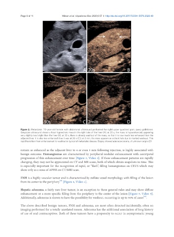
Page 6 of 11                                           Wilson et al. Hepatoma Res 2020;6:57  I  http://dx.doi.org/10.20517/2394-5079.2020.48


               Figure 2. Metastasis. 70-year-old female with abdominal ultrasound performed for right upper quadrant pain, query gallstones.
               Greyscale ultrasound shows a focal hypoechoic mass in the right lobe of the liver (A); at 20 s, the mass is hypoenhanced, appearing
               very slightly less bright than the liver (B); at 30 s, there is already washout of the mass, so that it is now much less enhanced than the
               adjacent liver. It is also less enhanced than it was at 20 s (C); at 2 min, the mass appears as a black hole due to marked washout. This
               rapid transition from enhancement to washout is typical of metastatic disease. Biopsy showed adenocarcinoma, of unknown origin (D)

               remain as enhanced as the adjacent liver to 4 or even 5 min following injection, is highly associated with
               benign outcome. Hemangiomas are characterized by peripheral nodular enhancement with centripetal
               progression of this enhancement over time [Figure 3, Video 3]. If these enhancement patterns are rapidly
               changing, they may not be appreciated on CT and MR scans, both of which obtain snapshots in time. This
               is especially important for the recognition of rapid, or “flash”, filling hemangiomas on CEUS which may
               show only as a zone of APHE on CT/MRI scan.


               FNH is a highly vascular tumor and is characterized by stellate vessel morphology with filling of the lesion
                                          [24]
               from its center to the periphery  [Figure 4, Video 4].

               Hepatic adenoma, a fairly rare liver tumor, is an exception to these general rules and may show diffuse
               enhancement or a more specific filling from the periphery to the center of the lesion [Figure 5, Video 5].
                                                                                                   [25]
               Additionally, adenoma is shown to have the possibility for washout, occurring in up to 50% of cases .
               The above described benign tumors, FNH and adenoma, are most often detected incidentally, often on
               imaging performed for a totally unrelated reason. Adenoma has the additional association of long history
               of use of oral contraceptives. Both of these tumors have a propensity to occur in asymptomatic young