
Page 93 - Read Online
P. 93
Wilson et al. Hepatoma Res 2020;6:57 I http://dx.doi.org/10.20517/2394-5079.2020.48 Page 7 of 11
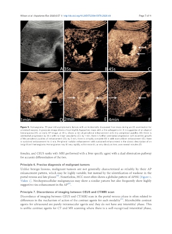
Figure 3. Hemangioma. 39-year-old asymptomatic female with an incidentally discovered liver mass during an US examination for
unrelated reasons. A greyscale image shows a focal slightly hypoechoic mass with a thin echogenic rim. It is suggestive of an atypical
hemangioma (A); an early AP image, at 35 s, shows a rim of peripheral enhancement with tiny peripheral puddles (B); there is
centripetal progression by 45 s with no linear vascularity (C); by 1 min, there is further centripetal progression with eccentric growth
of the peripheral puddles of enhancement (D); by 2 min, there is virtually complete fill in with non-uniform enhancement (E); there
is sustained enhancement to 4 min. Peripheral nodular enhancement with sustained enhancement is the classic description of an
insignificant hemangioma. Hemangiomas may fill very rapidly, within seconds, or very slowly as here, over several minutes (F)
females, and CEUS ranks with MRI performed with a liver specific agent with a dual elimination pathway
for accurate differentiation of the two.
Principle 6. Precise diagnosis of malignant tumors
Unlike benign lesions, malignant tumors are not generally characterized as reliably by their AP
enhancement pattern, which may be highly variable, but instead by the identification of washout in the
[26]
portal venous and late phases . Nonetheless, HCC most often shows a globular pattern of APHE [Figure 1,
Video 1]. Nonhepatocellular malignancies may show a similar pattern but also frequently show highly
[27]
suggestive rim enhancement in the AP .
Principle 7. Discordance of imaging between CEUS and CT/MRI scan
Discordance of imaging between CEUS and CT/MRI scan in the portal venous phase is often related to
[11]
differences in the mechanism of action of the contrast agents for each modality . Microbubble contrast
agents for ultrasound are purely intravascular agents and they do not have any interstitial phase. This
is unlike contrast agents for CT and MR scanning where there is a well-recognized interstitial phase,