
Page 100 - Read Online
P. 100
Brunsing et al. Hepatoma Res 2020;6:59 I http://dx.doi.org/10.20517/2394-5079.2020.50 Page 3 of 16
A
B
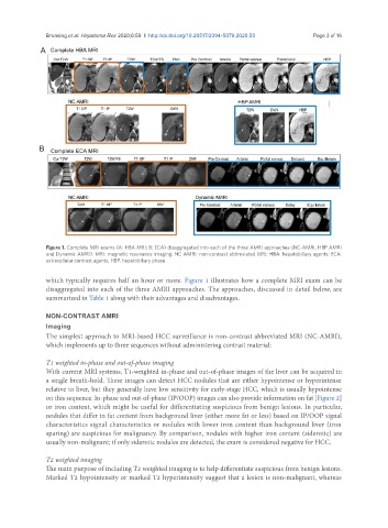
Figure 1. Complete MRI exams (A: HBA MRI; B: ECA) disaggregated into each of the three AMRI approaches (NC-AMRI, HBP AMRI
and Dynamic AMRI). MRI: magnetic resonance imaging; NC AMRI: non-contrast abbreviated MRI; HBA: hepatobiliary agents; ECA:
extracellular contrast agents; HBP: hepatobiliary phase
which typically requires half an hour or more. Figure 1 illustrates how a complete MRI exam can be
disaggregated into each of the three AMRI approaches. The approaches, discussed in detail below, are
summarized in Table 1 along with their advantages and disadvantages.
NON-CONTRAST AMRI
Imaging
The simplest approach to MRI-based HCC surveillance is non-contrast abbreviated MRI (NC-AMRI),
which implements up to three sequences without administering contrast material:
T1 weighted in-phase and out-of-phase imaging
With current MRI systems, T1-weighted in-phase and out-of-phase images of the liver can be acquired in
a single breath-hold. These images can detect HCC nodules that are either hypointense or hyperintense
relative to liver, but they generally have low sensitivity for early-stage HCC, which is usually hypointense
on this sequence. In-phase and out-of-phase (IP/OOP) images can also provide information on fat [Figure 2]
or iron content, which might be useful for differentiating suspicious from benign lesions. In particular,
nodules that differ in fat content from background liver (either more fat or less) based on IP/OOP signal
characteristics signal characteristics or nodules with lower iron content than background liver (iron
sparing) are suspicious for malignancy. By comparison, nodules with higher iron content (siderotic) are
usually non-malignant; if only siderotic nodules are detected, the exam is considered negative for HCC.
T2 weighted imaging
The main purpose of including T2 weighted imaging is to help differentiate suspicious from benign lesions.
Marked T2 hypointensity or marked T2 hyperintensity suggest that a lesion is non-malignant, whereas