
Page 97 - Read Online
P. 97
Page 8 of 17 Rastogi. Hepatoma Res 2020;6:47 I http://dx.doi.org/10.20517/2394-5079.2020.35
Figure 4. HCC in a non-cirrhotic background: HCC with pseudoglandular pattern in a background with F0-F1 fibrosis (A, HE stain;
B Masson Trichrome stain), HCC with microtrabecular pattern in F0 fibrosis (C, HE stain), steatohepatitic HCC with F3 fibrosis in
background liver (D, MT stain). HCC: hepatocellular carcinoma
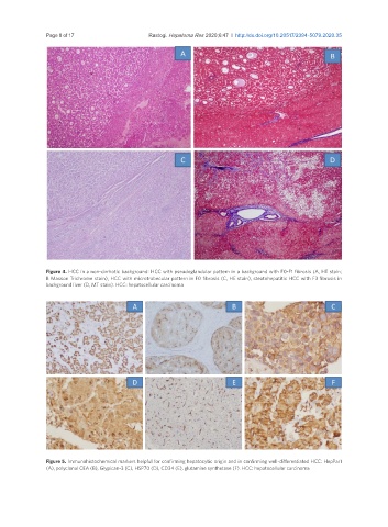
Figure 5. Immunohistochemical markers helpful for confirming hepatocytic origin and in confirming well-differentiated HCC: HepPar1
(A), polyclonal CEA (B), Glypican-3 (C), HSP70 (D), CD34 (E), glutamine synthetase (F). HCC: hepatocellular carcinoma