
Page 99 - Read Online
P. 99
Page 10 of 17 Rastogi. Hepatoma Res 2020;6:47 I http://dx.doi.org/10.20517/2394-5079.2020.35
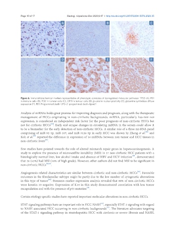
Figure 6. Immunohistochemical markers representative of phenotypic correlates of dysregulated molecular pathways: TP53 (A), PD1
in immune cells (B), PDL1 in tumour cells (C), CK19 in tumour cells (D), β-catenin nuclear positivity (E), glutamine synthetase diffuse
expression (F). PD1: Programmed death-1; PD-L1: programmed death-ligand 1
Analysis of miRNAs holds great promise for improving diagnosis and prognosis, along with the therapeutic
management of HCCs originating in non-cirrhotic backgrounds. miRNA, particularly hsa-mir-149
expression, is considered an independent risk factor for the poor prognosis of non-cirrhotic HCCs but
[85]
not for cirrhotic HCCs . Early and unique changes in circulating miRNA in the serum could allow it
to be a biomarker for the early detection of non-cirrhotic HCCs. A similar role of a three mi-RNA panel
[86]
comprising of miR-92-3p, miR-107, and miR-3126-5p in early HCC was shown by Zhang et al. and
Koh et al. reported the difference in expression of 16 miRNAs between non-tumor and HCC tissues in
[87]
[87]
non-cirrhotic livers .
Few studies have pointed towards the role of altered mismatch repair genes in hepatocarcinogenesis. A
study to explore the presence of microsatellite instability (MSI) in 37 non-cirrhotic HCC patients with a
[88]
histologically normal liver, low alcohol intake and absence of HBV and HCV infection , demonstrated
that 26 (43%) had MSI (16% of high grade). However, other authors did not find MSI to be significant in
non-cirrhotic HCCs [88,89] .
[90]
Angiogenesis related characteristics are similar between cirrhotic and non-cirrhotic HCCs . Favorable
outcomes in the fibrolamellar subtype might be partly due to the low number of cytogenetic aberrations
[91]
in this type of tumor . Stemness marker expression analysis revealed that 88% of non-cirrhotic HCCs
were keratin-19 negative. Expression of K19 in this study demonstrated correlation with less tumor
[39]
encapsulation and with the presence of p53 mutation .
Certain etiology specific studies have reported important molecular alterations in non-cirrhotic HCCs.
[92]
STAT signaling pathways have an important role in HCC-NASH , especially STAT-3 signaling with regard
to NASH associated HCC occurring in non-cirrhotic backgrounds . The literature advocates targeting
[93]
of the STAT-1 signaling pathway in steatohepatitic HCC with cirrhosis or severe fibrosis and NASH,