
Page 93 - Read Online
P. 93
Page 4 of 17 Rastogi. Hepatoma Res 2020;6:47 I http://dx.doi.org/10.20517/2394-5079.2020.35
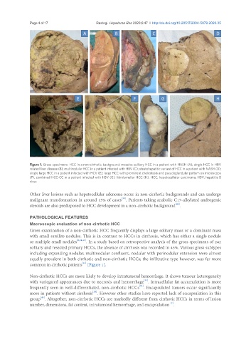
Figure 1. Gross specimens: HCC in a non-cirrhotic background: massive solitary HCC in a patient with NASH (A); single HCC in HBV
related liver disease (B); multinodular HCC in a patient infected with HBV (C); steatohepatitic variant of HCC in a patient with NASH (D);
single large HCC in a patient infected with HCV (E); large HCC with prominent cholestasis and pseudoglandular pattern on microscopy
(F); combined HCC-CC in a patient infected with HBV (G); fibrolamellar HCC (H). HCC: hepatocellular carcinoma; HBV: hepatitis B
virus
Other liver lesions such as hepatocellular adenoma occur in non-cirrhotic backgrounds and can undergo
[39]
malignant transformation in around 15% of cases . Patients taking anabolic C17-alkylated androgenic
[40]
steroids are also predisposed to HCC development in a non-cirrhotic background .
PATHOLOGICAL FEATURES
Macroscopic evaluation of non-cirrhotic HCC
Gross examination of a non-cirrhotic HCC frequently displays a large solitary mass or a dominant mass
with small satellite nodules. This is in contrast to HCCs in cirrhosis, which has either a single nodule
or multiple small nodules [6,24,41] . In a study based on retrospective analysis of the gross specimens of 242
solitary and resected primary HCCs, the absence of cirrhosis was recorded in 45%. Various gross subtypes
including expanding nodular, multinodular confluent, nodular with perinodular extension were almost
equally prevalent in both cirrhotic and non-cirrhotic HCCs; the infiltrative type however, was far more
common in cirrhotic patients [Figure 1].
[42]
Non-cirrhotic HCCs are more likely to develop intratumoral hemorrhage. It shows tumour heterogeneity
[24]
with variegated appearances due to necrosis and hemorrhage . Intracellular fat accumulation is more
[24]
frequently seen in well-differentiated, non-cirrhotic HCCs . Encapsulated tumors occur significantly
[12]
more in patients without cirrhosis . However other studies have reported lack of encapsulation in this
group . Altogether, non-cirrhotic HCCs are markedly different from cirrhotic HCCs in terms of lesion
[43]
[7]
number, dimensions, fat content, intratumoral hemorrhage, and encapsulation .