
Page 217 - Read Online
P. 217
Loh et al. Extracell Vesicles Circ Nucleic Acids 2023;4:568-87 https://dx.doi.org/10.20517/evcna.2023.34 Page 578
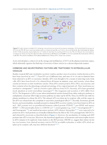
Figure 7. F-actin network controls DCV tethering, transport and secretion at the plasma membrane. (A) F-actins block the access of
DCVs to the PM; (B) Actin-severing proteins such as scinderin and gelsolin cut F actins into small filaments, thus facilitating the release
of DCV. PI3K causes the depolymerization of F-actins to facilitate the docking of DCVs to the PM; (C) Myosin Va is activated by
2+
increased Ca levels during stimulated secretion and interacts with Rab27a and MyRIP on DCVs to facilitate the mobilization of DCVs
to the PM. PM: plasma membrane; PI3K: phosphatidylinositol 3 kinase; DCV: dense core vesicles.
Actin network plays a critical role in the storage and mobilization of DCVs at the plasma membrane region,
which ultimately regulates the final step of secretion of these vesicles in a calcium-dependent manner.
HORMONE AND NEUROTROPHIC FACTORS ARE TRAFFICKED TO EXTRACELLULAR
VESICLES
Besides classical RSP and constitutive secretory vesicles, another class of secretory vesicles known as EVs
have been described in 1987 . These EVs are of different sizes, and ones of 30-150 nm in diameter have
[106]
been referred to as sEVs or exosomes. Initially, sEVs were thought to be a means of removing waste from
cells. sEVs have been found to be released from all tissues in animals, yeast, and bacteria, where these
vesicles are known as outer membrane vesicles. These sEVs contain protein, RNA, DNA, and enzymes, and
their contents reflect the physiology and pathophysiology of the original parental cell. Their membranes are
enriched in tetraspanins and are therefore quite different from DCVs. Recently, sEVs have garnished
[107]
[108]
much attention as novel intercellular messengers . The biogenesis and secretion of sEVs differ from
DCVs. The biogenesis of sEVs occurs when intraluminal vesicles formed from either endocytic pathway or
endoplasmic reticulum (ER)/Golgi secretory pathway are taken up by MVBs. Then, the MVBs fuse with the
plasma membrane to release the sEVs. The released sEVs are taken up by recipient cells, and the cargos in
the sEVs are released into the cytoplasm to exert their physiological effects . Recently, hormones, growth
[109]
factors, and neurotrophins, normally present in classical RSP secretory vesicles, have been found in sEVs as
[110]
well. RSP proteins such as parathyroid hormone-related protein (PTHrP) , pro-BDNF and mature
[111]
BDNF , CPE/neurotrophic factor-α1 (CPE/NF-α1) , several granins (e.g., CGA , Secretogranin III ,
[112]
[113]
[114]
and Neurogranin ), and amyloid precursor protein (APP)-derived Aβ peptides [109,116] were reported to be
[115]
present in sEVs. Such proteins are normally trafficked from the RER to the TGN, packaged into RSP DCVs,
and released by exocytosis as described above [Figure 1]. However, the mechanism of routing such RSP
proteins into sEVs is unclear. Moreover, the functional significance of hormones and neurotrophic factors
in sEVs remains to be explored. Some studies on the RSP protein, CPE/NF-α1, suggest that release via the
two mechanisms, from classical secretory vesicles (DCVs) as soluble molecules, or within sEVs which are
taken up by recipient cells, may have different functions [112,117] .