
Page 60 - Read Online
P. 60
Page 8 of 21 Guo et al. Energy Mater. 2025, 5, 500041 https://dx.doi.org/10.20517/energymater.2024.214
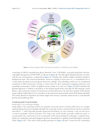
Figure 4. Schematic diagram of the in-situ/operando techniques in solid lithium polymer battery.
consisting of LiBOB, tetraethylene glycol dimethyl ether (TEGDME), and poly(vinylidene fluoride-
cohexafluoropropylene) (PVDF-HFP), as shown in Figure 6B. The SRS signal obtained directly correlates
with the ion concentration, as depicted in Figure 6C. Notably, this method exhibits minimal disruptive
background noise. The detection threshold, based on a signal-to-noise ratio of 1, is remarkably low at
10 mm under the given scanning speed and power settings. These findings underscore the exceptional
sensitivity of SRS microscopy, setting a robust basis for their subsequent imaging endeavors and
-2
quantitative assessments. Under an applied current density of 4.2 mA cm , Figure 6D and E visualizes the
gradual depletion of LiBOB in proximity to the lithium metal surface through 3D SRS imaging, which
shows a clear temporal evolution of the process. In their laboratory, the detection window of the Raman
setup could not satisfy direct Li-ion detection, so the researchers measured the anion BOB Raman intensity.
-
This study showed Li deposition has three stages: slow mossy Li growth, mixed mossy Li and dendrite
growth, and dendrite growth.
In-situ/operando X-ray technique
Small angle X-ray scattering technique
Small-angle X-ray scattering (SAXS) can quantify nanoscale electron density differences in a sample,
enabling detailed structural analysis through the scattering electron contrast between blocks of polymer
electrolytes [73-75] . SAXS typically provides structural information for sizes between 1 and 100 nm (up to
150 nm in partially ordered systems). The large-scale SAXS facility provides sufficient space to
accommodate the construction of an in-operando SAXS-electrochemical workstation coupled device.
Möhl et al. studied the nanoscale changes in polymer electrolytes in a capillary-based LIB using in-operando
SAXS [Figure 7A] . The radial SAXS profiles presented in Figure 7B and C reveal that the unannealed
[56]