
Page 32 - Read Online
P. 32
Soren et al. Cancer Drug Resist 2020;3:18-25 I http://dx.doi.org/10.20517/cdr.2019.106 Page 19
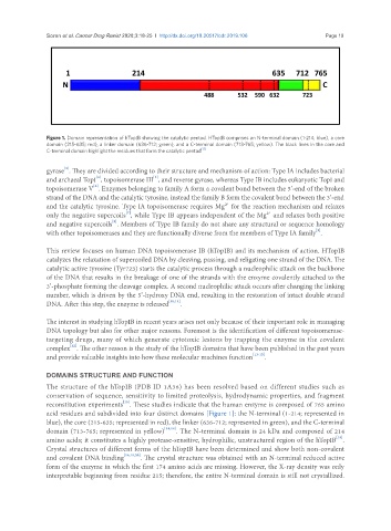
Figure 1. Domain representation of hTopIB showing the catalytic pentad. HTopIB comprises an N-terminal domain (1-214; blue), a core
domain (215-635; red), a linker domain (636-712; green), and a C-terminal domain (713-765; yellow). The black lines in the core and
C-terminal domain highlight the residues that form the catalytic pentad [17]
[3]
gyrase . They are divided according to their structure and mechanism of action: Type IA includes bacterial
[5]
[4]
and archaeal TopI , topoisomerase III , and reverse gyrase, whereas Type IB includes eukaryotic TopI and
[6]
topoisomerase V . Enzymes belonging to family A form a covalent bond between the 5’-end of the broken
strand of the DNA and the catalytic tyrosine, instead the family B form the covalent bond between the 3’-end
+
and the catalytic tyrosine. Type IA topoisomerase requires Mg² for the reaction mechanism and relaxes
[7]
+
only the negative supercoils , while Type IB appears independent of the Mg² and relaxes both positive
[8]
and negative supercoils . Members of Type IB family do not share any structural or sequence homology
[9]
with other topoisomerases and they are functionally diverse from the members of Type IA family .
This review focuses on human DNA topoisomerase IB (hTopIB) and its mechanism of action. HTopIB
catalyzes the relaxation of supercoiled DNA by cleaving, passing, and religating one strand of the DNA. The
catalytic active tyrosine (Tyr723) starts the catalytic process through a nucleophilic attack on the backbone
of the DNA that results in the breakage of one of the strands with the enzyme covalently attached to the
3’-phosphate forming the cleavage complex. A second nucleophilic attack occurs after changing the linking
number, which is driven by the 5’-hydroxy DNA end, resulting in the restoration of intact double strand
DNA. After this step, the enzyme is released [10,11] .
The interest in studying hTopIB in recent years arises not only because of their important role in managing
DNA topology but also for other major reasons. Foremost is the identification of different topoisomerase-
targeting drugs, many of which generate cytotoxic lesions by trapping the enzyme in the covalent
[12]
complex . The other reason is the study of the hTopIB domains that have been published in the past years
and provide valuable insights into how these molecular machines function [13-15] .
DOMAINS STRUCTURE AND FUNCTION
The structure of the hTopIB (PDB ID 1A36) has been resolved based on different studies such as
conservation of sequence, sensitivity to limited proteolysis, hydrodynamic properties, and fragment
[16]
reconstitution experiments . These studies indicate that the human enzyme is composed of 765 amino
acid residues and subdivided into four distinct domains [Figure 1]: the N-terminal (1-214; represented in
blue), the core (215-635; represented in red), the linker (636-712; represented in green), and the C-terminal
domain (713-765; represented in yellow) [14,18] . The N-terminal domain is 24 kDa and composed of 214
[18]
amino acids; it constitutes a highly protease-sensitive, hydrophilic, unstructured region of the hTopIB .
Crystal structures of different forms of the hTopIB have been determined and show both non-covalent
and covalent DNA binding [16,19,20] . The crystal structure was obtained with an N-terminal reduced active
form of the enzyme in which the first 174 amino acids are missing. However, the X-ray density was only
interpretable beginning from residue 215; therefore, the entire N-terminal domain is still not crystallized.