
Page 51 - Read Online
P. 51
Han et al. Cancer Drug Resist 2024;7:16 https://dx.doi.org/10.20517/cdr.2024.01 Page 13 of 25
*
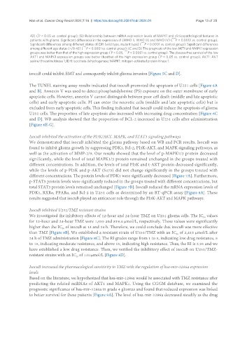
42) ( P < 0.05 vs. control group); (B) Relationship between mRNA expression levels of MAPK1 and clinicopathological features in
patients with glioma. Significant differences in the expression of (WHO II, WHO III, and WHO IV) ( **** P < 0.0001 vs. control group).
Significant differences among different states of IDH (wild type, mutant type) ( **** P < 0.0001 vs. control group); Significant differences
among different age status (</≥ 42) ( **** P < 0.0001 vs. control group); (C and D) The prognosis of the low AKT1 and MAPK1 expression
*
groups was better than that of the high expression group ( P < 0.05, **** P < 0.0001 vs. control group). The disease-free survival of the low
*
AKT1 and MAPK1 expression groups was better thanthat of the high expression group ( P < 0.05 vs. control group). AKT1: AKT
serine/threonine kinase 1; IDH: isocitrate dehydrogenase; MAPK1: mitogen-activated protein kinase 1.
isocuB could inhibit EMT and consequently inhibit glioma invasion [Figure 5C and D].
The TUNEL staining assay results indicated that isocuB promoted the apoptosis of U251 cells [Figure 6A
and B]. Annexin V was used to detect phosphatidylserine (PS) exposure on the outer membrane of early
apoptotic cells. However, annexin V cannot distinguish between poor cell death (middle and late apoptotic
cells) and early apoptotic cells. PI can enter the necrotic cells (middle and late apoptotic cells) but is
excluded from early apoptotic cells. This finding indicated that isocuB could induce the apoptosis of glioma
U251 cells. The proportion of late apoptosis also increased with increasing drug concentration [Figure 6C
and D]. WB analysis showed that the proportion of BCL-2 increased in U251 cells after administration
[Figure 6E-G].
IsocuB inhibited the activation of the PI3K/AKT, MAPK, and STAT3 signaling pathways
We demonstrated that isocuB inhibited the glioma pathway based on WB and PCR results. IsocuB was
found to inhibit glioma growth by suppressing PDK1, Bcl-2, PI3K-AKT, and MAPK signaling pathways, as
well as the activation of MMP-2/9. Our results showed that the level of p-MAPK1/3 protein decreased
significantly, while the level of total MAPK1/3 protein remained unchanged in the groups treated with
different concentrations. In addition, the levels of total PI3K and t-AKT protein decreased significantly,
while the levels of p-PI3K and p-AKT (S473) did not change significantly in the groups treated with
different concentrations. The protein levels of PDK1 were significantly decreased [Figure 7A]. Furthermore,
p-STAT3 protein levels were significantly reduced in the groups treated with different concentrations, but
total STAT3 protein levels remained unchanged [Figure 7B]. IsocuB reduced the mRNA expression levels of
PDK1, RXRα, PPARα, and Bcl-2 in U251 cells as determined by an RT-qPCR assay [Figure 8A]. These
results suggested that isocuB played an anticancer role through the PI3K-AKT and MAPK pathways.
IsocuB inhibited U251/TMZ resistant strains
We investigated the inhibitory effects of 12-hour and 24-hour TMZ on U251 glioma cells. The IC values
50
for 12-hour and 24-hour TMZ were 1,055 and 819.4 µmol/L, respectively. These values were significantly
higher than the IC of isocuB at 12 and 24 h. Therefore, we could conclude that isocuB was more effective
50
than TMZ [Figure 8B]. We established a resistant strain of U251/TMZ with an IC of 2,413 µmol/L after
50
24 h of TMZ administration [Figure 8C]. The RI grades range from 1 to 5, indicating low drug resistance, 5
to 15, indicating moderate resistance, and above 15, indicating high resistance. Thus, the RI is 2.95 and we
have established a low drug resistance. Then, we verified the inhibitory effect of isocuB on U251/TMZ-
resistant strains with an IC of 1.01µmol/L [Figure 8D].
50
IsocuB increased the pharmacological sensitivity to TMZ with the regulation of hsa-mir-1286a expression
levels
Based on the literature, we hypothesized that hsa-mir-1286a would be associated with TMZ resistance after
predicting the related miRNAs of AKT1 and MAPK1. Using the CGGM database, we examined the
prognostic significance of hsa-mir-1286a in grade 4 gliomas and found that reduced expression was linked
to better survival for these patients [Figure 9A]. The level of hsa-mir-1286a decreased steadily as the drug