
Page 143 - Read Online
P. 143
Page 10 of 18 Wang et al. Cancer Drug Resist. 2026;9:8
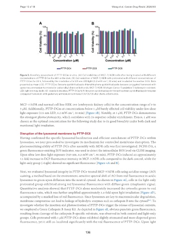
Figure 3. Biosafety assessment of PTTP-DCns in vitro. (A) Cell viabilities of MCF-7/ADR cells after being treated with different
concentrations of PTTP-DCns for 48 h in the dark; (B) Cell viabilities of MCF-7/ADR cells pretreated with different concentrations of
PTTP-DCns for 24 h, followed by the irradiation of a 525 nm LED light (0.2 mW·cm , 30 min) and incubated for another 24 h. Data
-2
presented as mean ± SD. PTTP-DCns: Benzene-pyridothiadiazole-thienothiophene-pyridothiadiazole-benzene conjugated framework with
quaternary ammonium-terminated n-carbon alkyl chains at both ends; MCF-7/ADR: Michigan Cancer Foundation-7/adriamycin-resistant;
LED: light emitting diode; SD: standard deviation; PTTP-DC4/6/8: benzene-pyridothiadiazole-thienothiophene-pyridothiadiazole-benzene
conjugated framework with quaternary ammonium-terminated C4/C6/C8 alkyl chains at both ends.
MCF-7/ADR and normal cell line HEK 293 (embryonic kidney cells) in the concentration range of 0 to
5 μM. Additionally, PTTP-DCns at concentrations below 1 μM barely affected cell viability under low-dose
light exposure (525 nm LED, 0.2 mW·cm , 30 min) [Figure 3B]. Notably, at 5 μM, PTTP-DC6 demonstrates
-2
the strongest photocytotoxicity, which correlates with its superior cellular enrichment. Hence, 1 μM was
chosen as the optimal concentration for the following study due to its good biosafety under both dark and
mentioned light irradiation.
Disruption of the lysosomal membrane by PTTP-DC6
Having confirmed the specific lysosomal localization and efficient enrichment of PTTP-DC6 within
lysosomes, we next proceeded to investigate its mechanism for controlled membrane disruption. The
photosensitizing ability of PTTP-DC6 after assembly with MDR cells was first investigated. DCFH-DA, a
green fluorescence-emitting ROS indicator, was used to detect the intracellular ROS level via CLSM imaging.
Upon ultra-low dose light exposure (525 nm, 0.2 mW·cm , 30 min), PTTP-DC6 induced an approximately
-2
12-fold increase in DCF fluorescence intensity in MCF-7/ADR cells compared to the dark control, while the
light-only group (+Light) showed no significant fluorescence [Figure 4A and B].
Next, we evaluated lysosomal integrity in PTTP-DC6-treated MCF-7/ADR cells using acridine orange (AO)
staining, a method based on the environment-sensitive spectral shift of AO from red fluorescence in acidic
lysosomes to green upon diffusion into the neutral cytosol. As shown in Figure 4C, cells in the light-only
pretreated group exhibited strong red lysosomal fluorescence with diffuse green cytoplasmic signal.
Quantitative analysis showed that PTTP-DC6 alone moderately increased the cytosolic green-to-red
fluorescence ratio, which was further amplified approximately 2.4-fold upon light irradiation [Figure 4D],
accompanied by a marked loss of red fluorescence. Since lysosomes are key to macromolecular degradation,
membrane compromise can lead to leakage of hydrolytic enzymes such as cathepsin B into the cytosol . To
[36]
investigate whether the insertion and photoactivation of PTTP-DC6 trigger the release of lysosomal contents,
we employed a Green Cathepsin B Assay Kit. As depicted in Figure 4E, obvious punctate green fluorescence,
resulting from cleavage of the cathepsin B-specific substrate, was observed in both control and light-only
groups. Cells pretreated with 1 μM PTTP-DC6 alone exhibited slightly attenuated and more dispersed green
fluorescence, yet it still co-localized significantly with the red fluorescence of PTTP-DC6. Upon light
136