
Page 76 - Read Online
P. 76
Kimbowa et al. Art Int Surg 2024;4:149-69 https://dx.doi.org/10.20517/ais.2024.20 Page 165
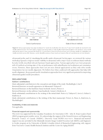
Figure 4. Various approaches have been developed for needle tip localization (A), but only 7 report both needle tip localization and
needle orientation error (B). Until 2022, progress was being made in reducing the needle tip localization error; however, most recent
approaches seem to have reversed this trend (A). Current state-of-the-art methods achieve sub-millimeter needle tip localization and
needle orientation errors (B). However, caution should be taken as these results are based on the self-reported errors on local datasets.
ultrasound probe; and (2) visualizing the needle under ultrasound. In this paper, we reviewed the various
methods proposed to improve needle visibility in ultrasound with a major focus on software-based methods
but also briefly described relevant hardware-based approaches. Various approaches have been proposed,
with AI methods achieving state-of-the-art performance with submillimeter tip localization and orientation
errors. However, these approaches have not yet seen clinical applications as they only solve needle
visualization but not needle alignment. We believe that when combined with approaches that help with
needle alignment, the proposed needle visualization approaches have very significant potential in improving
ultrasound-guided needle procedures.
DECLARATIONS
Authors’ contributions
Made substantial contributions to the conception and design of the study: Hacihaliloglu I, Liu D
Reviewed literature on needle appearance in ultrasound: Tadayon P
Reviewed literature on the hardware-based methods: Arora I, Pieters A
Reviewed literature on the software-based methods: Gulam S, Kimbowa A
Made substantial contributions to the writing of the initial draft: Pieters A, Tadayon P, Arora I, Gulam S,
Kimbowa A
Made substantial contributions to the writing of the final manuscript: Pieters A, Pinos A, Kimbowa A,
Hacihaliloglu I
Availability of data and materials
Not applicable.
Financial support and sponsorship
This work was supported by the Canadian Foundation for Innovation (CFI) - John R. Evans Leaders Fund
(JELF) program grant number 42816. We acknowledge the support of the Natural Sciences and Engineering
Research Council of Canada (NSERC). Discovery Grant RGPIN-2023-03575. Financial and material
support was also provided by the Minimally Invasive Image Guided Procedure Lab (MIIPs), and Engineers
in Scrubs program at the University of British Columbia School of Biomedical Engineering, with support
from Vancouver Coastal Health Research Institute and the Centre for Aging SMART.