
Page 74 - Read Online
P. 74
Page 164 Kimbowa et al. Art Int Surg 2024;4:149-69 https://dx.doi.org/10.20517/ais.2024.20
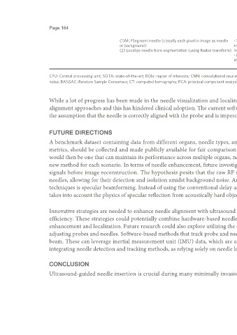
(1)MLPSegment needle (classify each pixel in image as needle - Segmentation method could be used to - Requires beam steering, assumes needle orientation is
or background) enhance needle visualization even without known a priori and is aligned with the selected beam steer
(2) Localize needle from segmentation (using Radon transform) localization angle
- Method robust with weak assumptions - Evaluated on data from 18-G needles. May not work for
about needle orientation thinner needles with non-distinctive appearance
- Only in-plane needle insertion
CPU: Central processing unit; SOTA: state-of-the-art; ROIs: region of interests; CNN: convolutional neural network; LSTM: long short-term memory; DBSCAN: density-based spatial clustering of applications with
noise; RANSAC: Random Sample Consensus; CT: computed tomography; PCA: principal component analysis; GPU: graphics processing unit; MLP: multi-layer perceptron.
While a lot of progress has been made in the needle visualization and localization approaches, little progress has been made in developing effective needle
alignment approaches and this has hindered clinical adoption. The current software-based methods alone cannot solve both challenges and they greatly rely on
the assumption that the needle is correctly aligned with the probe and is imperceptible to the naked eye.
FUTURE DIRECTIONS
A benchmark dataset containing data from different organs, needle types, and ultrasound scanners from multiple centers, with clearly defined evaluation
metrics, should be collected and made publicly available for fair comparison of any proposed software-based methods. An ideal needle detection method
would then be one that can maintain its performance across multiple organs, needle types, and ultrasound devices used, as it is not feasible to always develop a
new method for each scenario. In terms of needle enhancement, future investigations could focus on directly detecting needles from raw radio frequency (RF)
signals before image reconstruction. The hypothesis posits that the raw RF signal, being rich in information, may contain distinct patterns indicative of
needles, allowing for their detection and isolation amidst background noise. Another exciting avenue for future research on software-based image acquisition
techniques is specular beamforming. Instead of using the conventional delay-and-sum beamforming that assumes scattered reflection, specular beamforming
takes into account the physics of specular reflection from acoustically hard objects such as needles to improve their visibility in ultrasound .
[95]
Innovative strategies are needed to enhance needle alignment with ultrasound probes, particularly for inexperienced clinicians, while maintaining procedural
efficiency. These strategies could potentially combine hardware-based needle alignment approaches with software-based methods for needle visualization
enhancement and localization. Future research could also explore utilizing the entirety of video information, mirroring the approach taken by clinicians when
adjusting probes and needles. Software-based methods that track probe and needle motion can be developed to improve needle alignment with the ultrasound
beam. These can leverage inertial measurement unit (IMU) data, which are available in some ultrasound devices . Additionally, it is crucial to investigate
[96]
integrating needle detection and tracking methods, as relying solely on needle localization methods may not fully address the challenge of needle alignment .
[87]
CONCLUSION
Ultrasound-guided needle insertion is crucial during many minimally invasive procedures but faces two major challenges: (1) aligning the needle with the