
Page 460 - Read Online
P. 460
Page 6 of 7 Sengupta et al. Vessel Plus 2020;4:40 I http://dx.doi.org/10.20517/2574-1209.2020.55
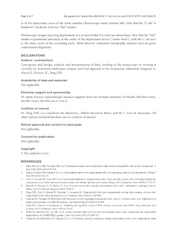
or in the inner/outer curve of the aortic annulus (fluoroscopic insets, bottom left). Note that the “C-tab” is
o
loaded 90 clockwise from the “Hat” marker.
Fluoroscopic images depicting deployment of a 26 mm Evolut Pro valve are shown here. Note that the “Hat”
marker is positioned anteriorly in the center of the deployment device (“center front”), with the C-tab seen
at the inner curve of the ascending aorta. Multi-detector computed tomography analyses indicate good
commissural alignment.
DECLARATIONS
Authors’ contributions
Conception and design, analysis and interpretation of data; drafting of the manuscript or revising it
critically for important intellectual content; and final approval of the manuscript submitted: Sengupta A,
Alexis SL, Kovacic JC, Tang GHL
Availability of data and materials
Not applicable.
Financial support and sponsorship
Dr. Jason Kovacic acknowledges research support from the National Institutes of Health (R01HL130423,
R01HL135093, R01HL148167-01A1).
Conflicts of interest
Dr. Tang GHL is a consultant for Medtronic, Abbott Structural Heart, and W. L. Gore & Associates. All
other authors declared that there are no conflicts of interest.
Ethical approval and consent to participate
Not applicable.
Consent for publication
Not applicable.
Copyright
© The Author(s) 2020.
REFERENCES
1. Mack MJ, Leon MB, Thourani VH, et al. Transcatheter aortic-valve replacement with a balloon-expandable valve in low-risk patients. N
Engl J Med 2019;380:1695-705.
2. Popma JJ, Deeb GM, Yakubov SJ, et al. Transcatheter aortic-valve replacement with a self-expanding valve in low-risk patients. N Engl J
Med 2019;380:1706-15.
3. Fuchs A, Kofoed KF, Yoon SH, et al. Commissural alignment of bioprosthetic aortic valve and native aortic valve following surgical and
transcatheter aortic valve replacement and its impact on valvular function and coronary filling. JACC Cardiovasc Interv 2018;11:1733-43.
4. Buzzatti N, Romano V, De Backer O, et al. Coronary access after repeated transcatheter aortic valve implantation: a glimpse into the
future. JACC Cardiovasc Imaging 2020;13:508-15.
5. Tang GHL, Zaid S, Ahmad H, Undemir C, Lansman SL. Transcatheter valve neo-commissural overlap with coronary orifices after
transcatheter aortic valve replacement. Circ Cardiovasc Interv 2018;11:e007263.
6. Gunning PS, Vaughan TJ, McNamara LM. Simulation of self expanding transcatheter aortic valve in a realistic aortic root: implications of
deployment geometry on leaflet deformation. Ann Biomed Eng 2014;42:1989-2001.
7. Tang GHL, Kaneko T, Cavalcante JL. Predicting the feasibility of post-TAVR coronary access and redo TAVR: more unknowns than
knowns. JACC Cardiovasc Interv 2020;13:736-8.
8. Webb JG, Murdoch DJ, Alu MC, et al. 3-year outcomes after valve-in-valve transcatheter aortic valve replacement for degenerated
bioprostheses: the PARTNER 2 registry. J Am Coll Cardiol 2019;73:2647-55.
9. Lederman RJ, Babaliaros VC, Rogers T, et al. Preventing coronary obstruction during transcatheter aortic valve replacement: from