
Page 458 - Read Online
P. 458
Page 4 of 7 Sengupta et al. Vessel Plus 2020;4:40 I http://dx.doi.org/10.20517/2574-1209.2020.55
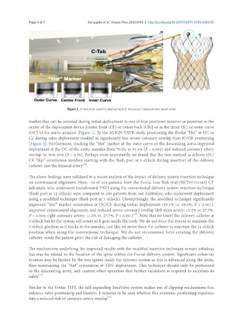
Figure 1. Orientation-specific deployment of the evolut transcatheter heart valve
marker that can be oriented during initial deployment in one of four positions: anterior or posterior in the
center of the deployment device [center front (CF) or center back (CB)] or in the inner (IC) or outer curve
(OC) of the aortic annulus [Figure 1]. In the ALIGN-TAVR study, positioning the Evolut “Hat” at OC or
CF during valve deployment resulted in significantly less severe coronary overlap than IC/CB positioning
[Figure 2]. Furthermore, tracking the “Hat” marker at the outer curve of the descending aorta improved
deployment at the OC of the aortic annulus from 70.2% to 91.6% (P = 0.002) and reduced coronary artery
overlap by 36%-60% (P < 0.05). Perhaps most importantly, we found that the best method to achieve OC/
CF “Hat” orientation involves starting with the flush port at 3 o’clock during insertion of the delivery
[12]
catheter into the femoral artery .
The above findings were validated in a recent analysis of the impact of delivery system insertion technique
on commissural alignment. Here, 154 of 249 patients from the Evolut Low Risk trial (NCT02701283) CT
sub-study who underwent transfemoral TAVI using the conventional delivery system insertion technique
(flush port at 12 o’clock) were compared to 240 patients from our institution who underwent deployment
using a modified technique (flush port at 3 o’clock). Unsurprisingly, the modified technique significantly
improved “Hat” marker orientation at OC/CF during initial deployment (93.1% vs. 69.6%, P < 0.001),
improved commissural alignment, and reduced severe coronary overlap (left main artery: 15.2% vs. 27.7%,
[17]
P = 0.004; right coronary artery: 11.8% vs. 27.7%, P < 0.001) . Note that we insert the delivery catheter at
3 o’clock but let the system self-rotate as it goes inside the body. We do not force the system to maintain the
3 o’clock position as it tracks to the annulus, just like we never force the catheter to maintain the 12 o’clock
position when using the conventional technique. We do not recommend force-rotating the delivery
catheter inside the patient given the risk of damaging the catheter.
The mechanisms underlying the improved results with the modified insertion technique remain nebulous
but may be related to the location of the spine within the Evolut delivery system. Significant columnar
rotation may be limited by the two spines inside the delivery system as this is advanced along the aorta,
thus maintaining the “Hat” orientation at THV deployment. This technique should only be performed
in the descending aorta, and caution other operators that further validation is required to ascertain its
[12]
safety .
Similar to the Evolut THV, the self-expanding JenaValve system makes use of clipping mechanisms that
enhance valve positioning and fixation. It remains to be seen whether this anatomic positioning translates
[14]
into a reduced risk of coronary artery overlap .