
Page 113 - Read Online
P. 113
Mathew et al. Vessel Plus 2020;4:11 I http://dx.doi.org/10.20517/2574-1209.2019.35 Page 9 of 15
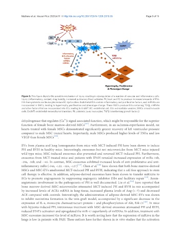
Figure 3. This figure depicts the possible mechanism of injury resulting in dysregulation of a number of vascular and inflammatory cells.
Injury (inflammatory, oxidant, drug toxicity, increased pulmonary flow) activates Plt, Leuk and EC to produce increased amounts of EVs.
EVs from platelets and leukocytes induce EC dysfunction. Endothelial EVs contain inflammatory and proliferative factors, and miRNAs are
incorporated in SMCs, leading to hypertrophy, proliferation and phenotype change. These SMCs produce EVs containing TGFβ, miRNAs
and other factors that are incorporated into ECs, leading to EndMT. EC: endothelial cell; EVs: extracellular vesicles; SMCs: smooth muscle
cells; EndMT: endothelial mesenchymal transition; Plt: platelets; Leuk: leukocytes; TGFβ: transforming growth factor β
2+
dehydrogenase that regulates (Ca )i signal associated function, which might be responsible for the superior
function of female bone marrow-derived MSCs [117] . Furthermore, in an ischemia-reperfusion model, rat
hearts treated with female MSCs demonstrated significantly greater recovery of left ventricular pressure
compared to male MSC treated hearts. Importantly, male MSCs produced higher levels of TNFα and less
VEGF than female MSCs [118] .
EVs from plasma and lung homogenates from mice with MCT-induced PH have been shown to induce
PH and RVH in healthy mice. Interestingly, exosomes but not microvesicles from MCT-mice injured
wild type mice; MSC-induced exosomes also prevented and reversed MCT-induced PH. Furthermore,
exosomes from MCT-treated mice and patients with IPAH revealed increased expression of miRs-19b,
-20a, -20b, and -145. In contrast, MSC-exosomes exhibited increased levels of anti-proliferative and anti-
inflammatory miRs (-34a, -122. -124, -127) [119] . Chen et al. [120] have shown that both bone marrow-derived
MSCs and MSC-EVs ameliorated MCT-induced PH and RVH, indicating that a cell free approach to stem
cell therapy is effective. In addition, adipose-derived exosomes have been shown to transfer miR125a to
ECs to promote angiogenesis by suppressing angiogenic inhibitor Dll4 and facilitate repair [121] . Renin-
angiotensin involvement in the pathogenesis of PH is well documented. Liu et al. [122] have reported that
bone marrow-derived MSC-microvesicles attenuated MCT-induced PH and RVH in rats accompanied
by increased levels of ACE2 mRNA in lung tissue, increased plasma levels of Ang-(1-7) and decreased
ACE compared with controls. Interestingly, the administration of adipose-derived MSC-EVs was shown
to inhibit neointima formation in the vein graft model, accompanied by a significant decrease in the
expression of IL-6, monocyte chemoattractant protein-1 and phosphorylation of Akt, Erk1/2 [123] . In mice
with hypoxia-induced PH, intravenous treatment with MSC-derived exosomes attenuated PH and RVH,
reduced STAT3 activation and upregulated the miR-17 superfamily of miRNAs. In addition, treatment with
MSC exosomes increased the level of miR204. It is worth noting here that the expression of miR204 in the
lungs is low in patients with PAH. These authors have further shown in in vitro studies that the activation